Nonepithelial Biliary Tumors
Michael P. Federle, MD, FACR
Key Facts
Imaging
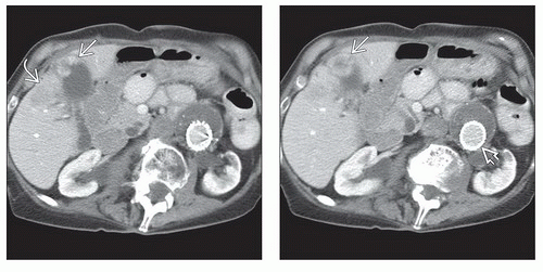
Nonepithelial tumors of the gallbladder (GB) and bile ducts are usually indistinguishable (by imaging) from primary GB carcinoma or cholangiocarcinoma, respectively
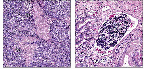
Distinction is made by histology and immunohistochemical characterization of tissue samples following resection
Distinction is important as prognosis and treatment are markedly different for various tumors
Arise in submucosal or smooth muscle layer of the biliary system
GB tumors are usually polypoid and may reach considerable size before symptoms develop
Biliary tumors are usually diagnosed while small due to early onset of symptoms (jaundice)
Top Differential Diagnoses
Gallbladder carcinoma
Gallstones are present; tumor is hypovascular
Cholangiocarcinoma
Cholangitis, primary sclerosing
Inflammatory pseudotumor, hepatobiliary
Pathology
Neuroendocrine tumor (NET) of biliary system
Well-differentiated NET (carcinoid)
Poorly differentiated NET (poor prognosis)
Sarcomas of biliary system
More common in bile ducts than GB
Granular cell tumor
Benign neural tumor (sometimes called schwannoma)
IMAGING
General Features
Best diagnostic clue
Nonepithelial tumors of the gallbladder (GB) and bile ducts are usually indistinguishable (by imaging) from primary GB carcinoma or cholangiocarcinoma, respectively
Distinction is made by histology and immunohistochemical characterization of tissue samples following resection
Distinction is important as prognosis and treatment are markedly different for various tumors
Location
Arise in submucosal or smooth muscle layer of the biliary system
Size
GB tumors are usually polypoid and may reach considerable size before symptoms develop
Biliary tumors are usually diagnosed while small due to early onset of symptoms (jaundice)
Stay updated, free articles. Join our Telegram channel

Full access? Get Clinical Tree


