Nonneoplastic Cysts
Arachnoid Cyst
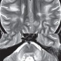
Arachnoid cysts are common, benign, CSF filled lesions. They represent 1% of all intracranial masses. In terms of etiology, they can be congenital, inflammatory, or post-traumatic. The most common location is the middle cranial fossa ( Fig. 1.140 ) with other characteristic locations including overlying the brain convexity, in the perimesencephalic cistern, and posterior to the cerebellum. In the middle cranial fossa, arachnoid cysts can be accompanied by hypogenesis of the temporal lobe. The vast majority of arachnoid cysts are asymptomatic, although symptoms due to mass effect can occur. On CT, communication of these lesions with the subarachnoid space can be demonstrated, with filling by intrathecal contrast on delayed scans. On MR, arachnoid cysts will demonstrate CSF signal intensity on all pulse sequences. Although the appearance of an arachnoid cyst is characteristic, a consideration of two other entities that show some similarity on imaging is likely warranted. Epidermoid tumors on all scans other than diffusion are relatively isointense to CSF, and like arachnoid cysts are space occupying masses. However, epidermoids are distinctive in having marked high signal intensity on diffusion weighted scans. Cystic neoplasms can be differentiated on the basis of demonstration of the cyst wall, abnormal contrast enhancement of a portion of the lesion, associated abnormal soft tissue, non-CSF signal intensity of the fluid, and accompanying vasogenic edema.

Related posts:

Stay updated, free articles. Join our Telegram channel

Full access? Get Clinical Tree


