Introduction
It is not unusual to encounter incidental findings during the course of a venous examination. Some of these findings may be the cause of extremity pain or swelling apart from venous disease. Recognition of the sonographic features associated with different pathologies is critical for accurate diagnosis. Many of these diagnoses are common, such as edema, hematoma, lymph nodes, or popliteal (Baker) cysts. Other findings are less common, but no less important, including abscess, joint effusion, adenopathy, benign or malignant tumor, and metastatic disease.
It is always important to discuss severity, timing, and location of pain and/or swelling with the patient during the study. Be sure to examine the areas of concern in addition to your standard protocol views. Not only does this improve your detection of important findings, it also allows your patient an opportunity to describe the reason for their visit.
Soft Tissue Edema
Lower extremity edema is a common cause of limb swelling and is associated with either elevated venous pressures or lymphatic obstruction (lymphedema), or both. Causes of increased hydrostatic pressure within the venous system include heart failure (HF; venous congestion), fluid overload, deep vein thrombosis (DVT), venous compression, and other causes of venous obstruction. These problems result in leg swelling and soft tissue edema.
There is a change in venous flow patterns in cases of heart failure. We expect to see phasic, nonpulsatile waveforms in the normal lower extremity veins. In right-sided heart failure, the waveforms are usually more pulsatile and typically bidirectional, with components seen above and below the baseline ( Fig. 22.1 ). As noted in Chapter 1 , causes of increased venous pressure such as heart failure or tricuspid insufficiency lead to increased transmission of cardiac phasic changes in pressure and blood flow to the peripheral veins of the upper and lower limbs. These changes result in increased pulsatility in the peripheral vein waveforms. Pulsatility in the venous system may resemble arterial pulsations because of the bidirectional pattern of blood flow. Comparison of the arterial and venous flow patterns is key to distinguishing pulsatile venous signals from arterial flow. On close inspection, the phases of the venous pattern are less periodic than the typical triphasic waveforms found in the arterial system. Other clues to the nature of the flow abnormality are the direction of flow and the communications with the superficial veins. Another distinguishing characteristic is the change in venous flow that occurs with the Valsalva maneuver. This does not occur on the arterial side.


Pulsatile venous waveforms from venous congestion are frequently mistaken for signals associated with arteriovenous fistula (AVF). Although blood flow patterns seen with both venous congestion and AVF will demonstrate increased pulsatility, there are major differences. The waveforms identified from both the arterial and venous sides of an AVF will show an arterialized pattern with a low-resistance (high-diastolic) component. Blood flow in the fistula will only be in one direction toward the venous or lower-pressure side without reversal of flow. This is very different from the typical biphasic (above and below the baseline) pattern seen with venous congestion.
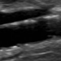
Gray-scale imaging may show a marbled, reticulated pattern when edema collects in the subcutaneous fat. Small collections of fluid may be seen when edema collects in the superficial tissues ( Fig. 22.2 ) and can be associated with significant soft tissue swelling. Attenuation of sound may occur that can restrict visualization of deep tissues and underlying vascular structures. A sound approach to this problem is to switch transducers to allow lower-frequency imaging. Changing from the usual 5 to 7 MHz linear array probe to a 2 to 5 MHz curved array transducer should allow deeper penetration of sound and improved visualization of the deeper areas of interest.

Bilateral leg swelling is a typical presentation for patients with venous congestion. It is also a common indication for venous sonography. Studies have shown that the likelihood of identifying DVT in patients with bilateral leg swelling without significant risk factors is low (≤5%). Conversely, Naidich and associates showed that 23% of patients with bilateral symptoms and risk factors had underlying DVT. In this group, 78% of patients had risk factors for DVT. DVT cannot be reliably diagnosed or excluded on clinical grounds alone.
Venous compression and venous obstruction are also causes of leg swelling and edema. The unexplained presence of edema, in association with abnormal waveforms should prompt an investigation of the proximal (or central) venous structures for thrombosis or compression.
Unlike the increased pulsatility seen with increased venous pressures and venous congestion, decreased phasicity is noted with venous obstruction or compression ( Fig. 22.3 ). Causes of central venous thrombosis include inferior vena cava and iliac vein DVT. Venous compression may be caused by lymphadenopathy, metastatic tumor, aneurysm, fluid collection, pelvic mass, gravid uterus, and an overdistended bladder.


Lymphedema
Lymphedema is also associated with leg swelling and can mimic DVT. Obstruction of the lymphatics due to malignancy, trauma, or surgery will cause extremity swelling and pain. Patients will typically present with unilateral or bilateral leg swelling. The appearance of lymphedema is indistinguishable from soft tissue edema associated with venous congestion. There may or may not be lymph node enlargement to suggest the etiology of the leg swelling.
- •
There are multiple nonvascular findings that can indicate the cause of leg pain and swelling apart from venous thrombosis.
- •
Many of the causes are common, including edema, hematoma, lymph nodes, and popliteal cysts.
- •
Ultrasound will not only identify many of the common causes of leg symptoms but also identify significant and unsuspected pathologies including abscesses, tumors, and metastatic disease.
- •
Lower extremity edema is a common cause of limb swelling and is associated with elevated venous pressures or lymphedema.
- •
In right heart failure, venous waveforms are usually more pulsatile and typically bidirectional, with components seen above and below the baseline.
- •
Studies have shown that the likelihood of identifying DVT in patients with bilateral leg swelling without significant risk factors is low (≤5%). Conversely, 23% of patients with bilateral symptoms and risk factors had underlying DVT.
- •
DVT cannot be reliably diagnosed or excluded on clinical grounds alone.
Hematoma
Another common cause of leg swelling and pain is soft tissue hematoma. These patients may have focal or diffuse swelling of the extremity. Look for a history of anticoagulation, which suggests the correct diagnosis. Check also for a history of trauma, vigorous exercise, or prior surgery or intervention.
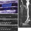
Sonographic examination of a palpable or focal hematoma may reveal a cystic, complex, or solid lesion that is typically found in the superficial tissues. The mass may initially have irregular borders and demonstrate areas of fluid and solid tissue depending on the amount of clot in the hematoma ( Fig. 22.4 ). Over time, the mass will retract and become better defined and ovoid. Hematoma may also be spread through fascial or muscle planes and seen as hypoechoic or isoechoic tissue. This may be very difficult to identify as there may be ill-defined borders and no significant difference in echogenicity from adjacent structures. Leg swelling may be the only clue to the soft tissue infiltration with hematoma. Magnetic resonance imaging (MRI) provides superior soft tissue contrast and displays the extent of a soft tissue hematoma.

Color and duplex Doppler will demonstrate absent flow signals within a hematoma. This is very helpful to distinguish a hematoma from other soft tissue masses such as lymph nodes or tumors. Hematoma may compress or displace adjacent vascular structures and demonstration of patent veins will allow exclusion of DVT. With significant compression by large hematomas, adjacent veins may be difficult to identify. Low-velocity blood flow below the level of compression may also be difficult to detect.
Muscle Injury
Leg pain from muscle injury is a common indication for venous ultrasound to exclude DVT. Muscle injury may result from a blow (contusion), from a penetrating injury, or from a muscle pull, which is a tearing of muscle bundles in response to vigorous exercise or abrupt straining. Tearing of muscle is accompanied initially by varying degrees of bleeding and, subsequently, by inflammation. Kim and colleagues reported that injured muscle initially is hyperechoic and either homogeneous or heterogeneous. Focal abnormalities may be related to hematoma and edema. By about 7 days after injury, the affected muscle is intermediate to low in echogenicity, and heterogeneous. These findings persist for at least 3 weeks after injury ( Fig. 22.5 ). Long-term follow-up information is not available. With a muscle pull, the area of injury may be quite focal and possibly fusiform in shape. Larger areas of abnormality may be seen with contusion.

Related posts:
 Anatomy of the Upper and Lower Extremity Arteries
 Ultrasound Assessment Following Endovascular Aortic Aneurysm Repair
 Ultrasound Assessment of Carotid Stenosis
 Assessment of Upper Extremity Arterial Disease
 Ultrasound Assessment Following Endovascular Aortic Aneurysm Repair
 Evaluation of Organ Transplants
Stay updated, free articles. Join our Telegram channel

Full access? Get Clinical Tree


