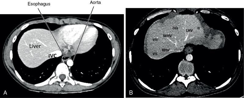
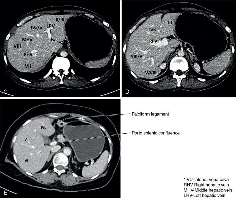
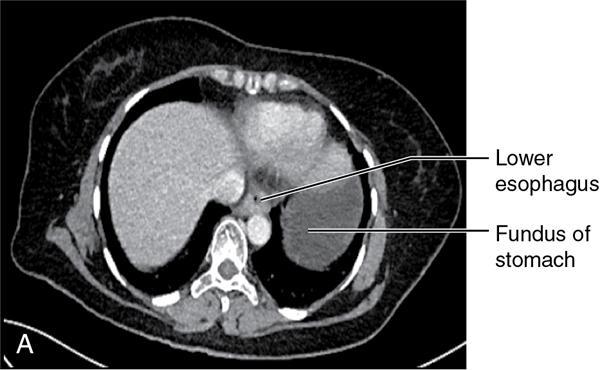
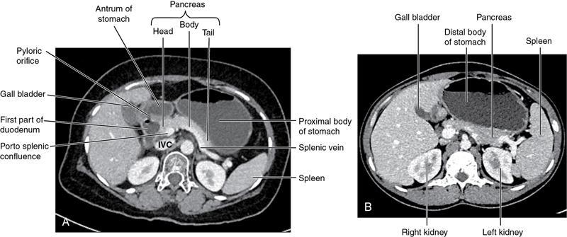
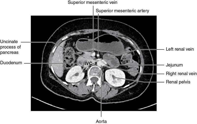
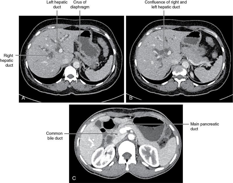
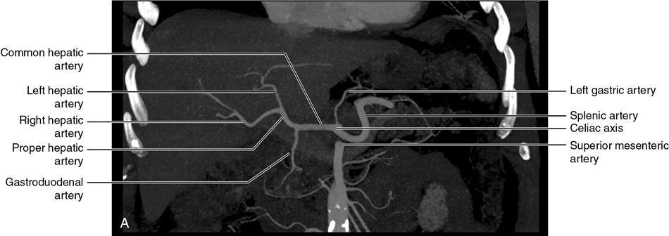
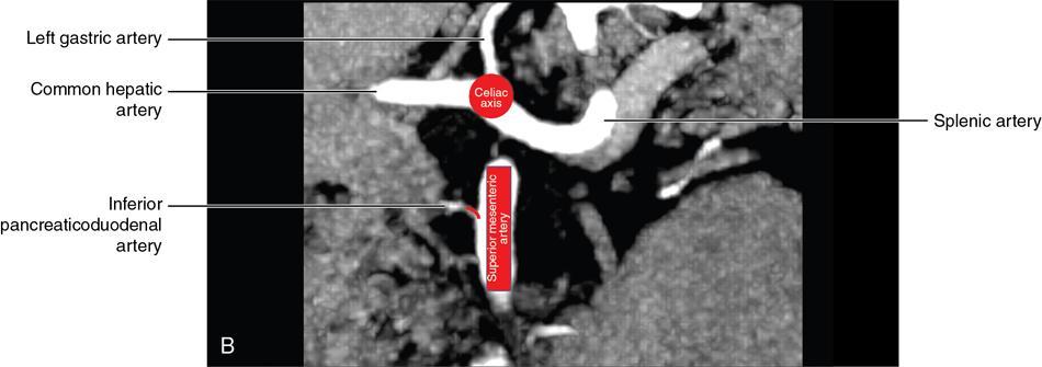
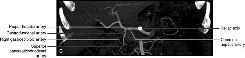
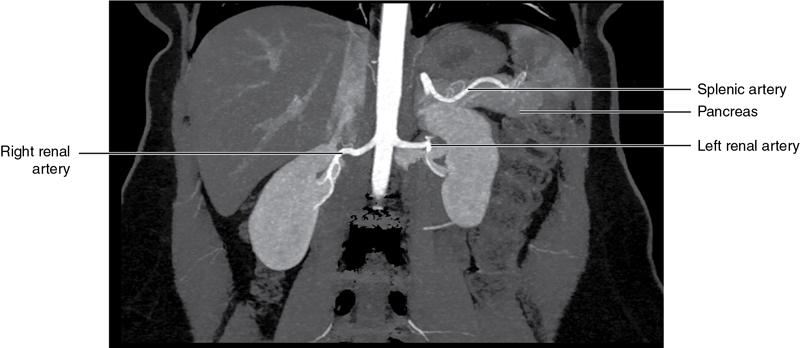
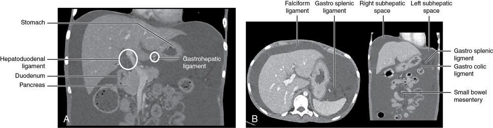
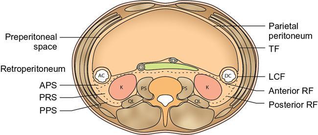
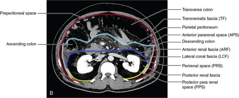
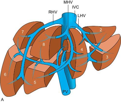
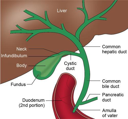
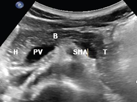
CROSS SECTIONAL ANATOMY OF ABDOMEN Satya Jha NORMAL ANATOMY OF ABDOMEN AND PELVIS Amandeep Singh The two major surfaces: The anterior and posterior layers of the coronary ligament converge on bare area (not covered by peritoneum). Its right and left margins form the right and left triangular ligaments. The right triangular ligament extends toward the diaphragm and separates right subphrenic space from right subhepatic space. The left triangular ligament gives tracts extending to the diaphragm and falciform ligament and does not compartmentalize the left subphrenic space. Ligamentum teres or the obliterated umblical vein is contained in falciform ligament which attaches the liver to anterior abdominal wall. The main portal vein, the proper hepatic artery and the common bile duct are contained within investing peritoneal folds of hepatoduodenal ligament at the porta hepatis (Fig. 7.2.2.1). Liver is divided into eight segments which are functionally independent and have their own vascular supply and biliary drainage. Arterial circulation: The branches of the hepatic artery accompanying the portal veins. Hepatic venous system: The right, middle, and left hepatic veins draining into IVC (Figs. 7.2.2.2 and 7.2.2.3). The gallbladder is a blind pear-shaped muscular membranous sac which is an embryologic derivative of the foregut, is a pouch lying along the undersurface of the liver. The gallbladder fossa is located in the plane of the interlobar fissure, which lies between the right and left hepatic lobes. Its major function is to store and concentrate bile which is produced by the liver. It measures approximately 4 cm in diameter when it is normally distended. Gallbladder is a smaller tubular structure in contracted state. The normal gallbladder wall thickness ranges from 1 to 3 mm. The gallbladder is divided into the fundus, body and neck. Infundibulum is present in the region of neck of the gallbladder, which is called the Hartmann pouch, where gallstones are usually impacted. Intrahepatic biliary radicles (IHBRs) scattered throughout the liver get confluent towards the hilum. They unite to form the right and left main hepatic ducts which further unite to form common hepatic duct (CHD) at the hilum. Common bile duct is formed by the union of cystic duct with common hepatic duct. The main pancreatic duct is joined with the common bile duct to form the ampulla of Vater at the major duodenal papilla (Figs. 7.2.2.4 and 7.2.2.5). Pancreas is located in anterior pararenal space of retroperitoneum anterior to perirenal (Gerota’s) fascia and posterior to parietal peritoneum. It is divided into head, uncinate process, neck, body and tail from right to left. Pancreas lies anterior to portal vein, which marks the point of transition between the body and neck. The region between head of pancreas and second and third parts of duodenum is known as the pancreatic groove. In postnephrectomy cases or with agenesis of kidney or ectopic kidney, pancreas moves posteriorly to partially fill in the empty renal fossa; its soft tissue density should not be mistaken for recurrent tumour. It is located in the pancreatic groove and is bounded superiorly by the duodenal bulb, laterally by second portion of duodenum, inferiorly by third portion of duodenum, medially by superior mesenteric vein and anterior to inferior vena cava. It is a wedge or wedge shaped lying posterior to superior mesenteric artery and vein. It is an imaginary junction between the head and body and lies directly over the junction of the splenic vein and superior mesenteric vein. It is located posterior to the lesser sac and anterior to the aorta, left adrenal gland, left kidney, and renal vessels and runs obliquely upward to the left of the superior mesenteric vessels. It is situated median to the colonic flexure and anterior to the left kidney. It is located in close proximity to the splenic hilum without a notable relation with the body of pancreas. It is seen anterior to the left kidney and median to the colonic flexure. The distal part of the tail passes between the peritoneal layers of the splenorenal ligament (Fig. 7.2.2.6 and 7.2.2.7).
7.2: Normal anatomy and normal variant
Liver

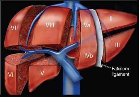
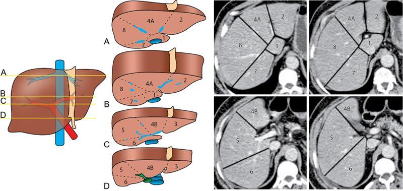
The bismuth and couinaud classification of liver
Couinaud
Traditional
Segment I
Caudate lobe
Segment II
Lateral superior segment of left lobe of liver
Segment III
Lateral inferior segment of left lobe of liver
Segment Iva
Medial superior segment of left lobe of liver
Segment IVb
Medial inferior segment of left lobe of liver
Segment V
Anterior inferior segment of right lobe of liver
Segment VI
Posterior inferior segment of right lobe of liver
Segment VII
Posterior superior segment of right lobe of liver
Segment VIII
Anterior superior segment of right lobe of liver
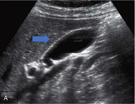
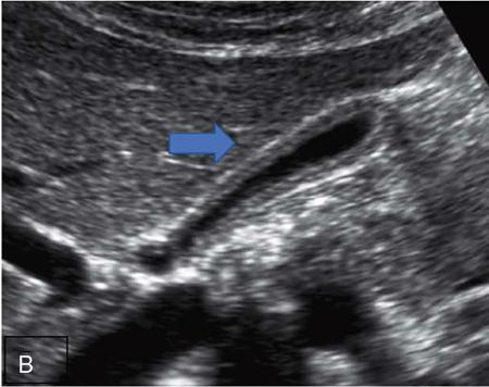
Gallbladder
Normal anatomy
Pancreas
Normal anatomy and relationships of pancreas
Head
Uncinate process
Neck
Body
Tail
Related posts:


Stay updated, free articles. Join our Telegram channel

Full access? Get Clinical Tree


