6 Normal Cerebrovascular Anatomy and Collateral Pathways
The vascular system of the human brain differs significantly, both anatomically and physiologically, from other organs in the body. Although it accounts for only 2% of the body weight, the brain receives 15% of the cardiac output and consumes 20% of the body’s oxygen supply in the basal state.1
Cerebral arteries are little influenced by sympathetic nerves, unlike other arteries, but they are markedly affected by chemical changes in the blood, especially changes in local carbon dioxide and oxygen levels.2,3
Vascular Anatomy
The blood supply for the central nervous system1,4,5 derives from the three great vessels arising from the aortic arch in the superior mediastinum—the brachiocephalic, the left common carotid, and the left subclavian arteries (Figure 6-1). The brachiocephalic artery travels upward, slightly posterior from the arch to the right of the neck for its 4- to 5-cm length, dividing into the right common carotid artery and the right subclavian artery at the upper border of the right sternoclavicular junction. The left common carotid artery ascends from the arch and passes beneath the left sternoclavicular joint. Neither common carotid has collateral branches, but each divides into the internal and external carotid arteries at the level of the upper border of the thyroid cartilage.
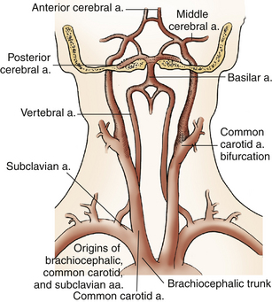
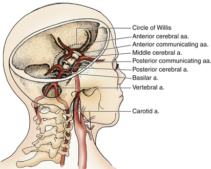
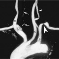
The internal carotids supply most of the anterior circulation to the cerebrum (Figure 6-2). In their cervical portion, the internal carotid arteries may be relatively straight or may take a tortuous course as they travel to the base of the skull. With the exception of very rare anomalies, there are no branches of the internal carotid arteries in the neck. As they proceed intracranially, the internal carotid arteries give rise to the caroticotympanic branches in the petrous bone, the meningohypophyseal branches in the cavernous sinus region, and the ophthalmic arteries immediately distal to the cavernous sinus. Eight millimeters beyond the clinoid process, within the dura mater, the internal carotid arteries give rise to the posterior communicating arteries, which join with the posterior cerebral arteries. Further cephalad, the internal carotid arteries divide into the middle and anterior cerebral arteries and give rise posteriorly to the anterior choroidal arteries.
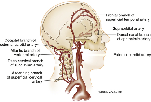
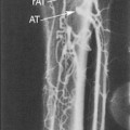
The external carotid arteries normally supply no blood to the brain. However, several of their branches can become important collateral pathways if occlusion occurs in the internal carotid or vertebral arteries. The branches of the external carotid artery are the ascending pharyngeal, the superior thyroid, the lingual, the external maxillary, the occipital, the facial, the posterior auricular, the internal maxillary, the transverse facial, and the superficial temporal arteries. The external carotid branches most vital to collateral circulation are those in communication with the ophthalmic artery and those that interconnect between the muscular branches of the occipital and vertebral arteries (Figure 6-3).
The posterior circulation to the brain is supplied in large part by the vertebral arteries. They arise from the subclavian arteries. The vertebral arteries lie within the foramina transversarium of the upper cervical vertebrae and wind anteriorly into the subarachnoid space at the side of the medulla oblongata at the level of the atlanto-occipital interspace. They proceed cephalad and anteriorly until they reach the pontomedullary level, where they join to form the basilar artery. Four branches arise from the basilar artery as it courses upward before dividing into the posterior cerebral arteries. Branches of the basilar artery supply the entire pons and the superior and anterior aspects of the cerebellum. Branches of the vertebral arteries supply the medulla and the interior surface of the cerebellum.
The cerebral branches of the internal carotid and vertebral arteries are joined at the base of the brain by an arterial circle known as the circle of Willis, the most important element of the intracranial collateral circulation and also a common site of aneurysm formation. It is a hexagonal arrangement of arteries composed of the anterior, middle, and posterior cerebral arteries, which are joined together by the anterior and posterior communicating arteries (see Figure 6-2). Under normal circumstances, there is little mixing of blood through the communicating arteries. However, in instances of arterial occlusion of the carotid or vertebrobasilar arteries, this circle opens to function as a vital collateral pathway (see later).
Component arteries of the circle of Willis can vary greatly in size, and there are at least nine congenital variations in the structure of the circle (Figure 6-4
Related posts:
 Risk Factors and the Role of Ultrasound in the Management of Extremity Venous Disease
 Ultrasound Assessment of Lower Extremity Arteries
 Ultrasound Assessment of the Vertebral Arteries
 Arterial Anatomy of the Extremities
 Duplex Ultrasound Evaluation of the Uterus and Ovaries
 Extremity Venous Anatomy and Technique for Ultrasound Examination
Stay updated, free articles. Join our Telegram channel

Full access? Get Clinical Tree


