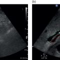
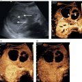
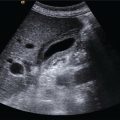
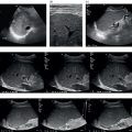
Adrian K.P. Lim1,2, Ivica Grgurevic3, and Matteo Rosselli4,5 1 Department of Imaging, Imperial College Healthcare NHS Trust, London, UK 2 Department of Metabolism, Digestion and Reproduction, Imperial College London, UK 3 Department of Gastroenterology, Hepatology and Clinical Nutrition, University Hospital Dubrava, University of Zagreb School of Medicine and Faculty of Pharmacy and Biochemistry, Zagreb, Croatia 4 Department of Internal Medicine, San Giuseppe Hospital, USL Toscana Centro, Empoli, Italy 5 Division of Medicine, Institute for Liver and Digestive Health, University College London, Royal Free Hospital, London, UK Ultrasound is considered one of the first‐line diagnostic tests for liver imaging. Based on its findings, decisions that have a direct impact on the patient pathway are made, leading to further investigations, follow‐up, or discharge. Along these lines, it should be kept in mind that greyscale ultrasound images lay out an anatomical and structural view of the liver, and therefore it is important to have a significant knowledge of abdominal anatomy and to ensure that the whole liver is assessed thoroughly. This chapter provides an overview of normal liver anatomy and its sonographic transposition, as well as a practical guide on how to start scanning the liver, with probe position and obtaining the standard recorded ultrasound images that should form the backbone of any liver ultrasound scan. A guide on how to report on a liver ultrasound study is also provided. The liver is the largest parenchymal organ in the abdominal cavity. It is located below the diaphragm, extending from the right hypochondrium to the epigastrium, usually reaching the left subcostal edge (Figure 3.1). It has a smooth, dome‐shaped diaphragmatic surface and a visceral, more irregular one, moulded by the adjacent organs and indented by the left, right, and interlobar fissures (Figure 3.2). Normally during respiration, the liver moves following the diaphragm. This movement is important and can be increased with deep inspiration to optimise liver visualisation during ultrasound imaging in a subcostal view. The magnitude of these excursions depends on the individual’s lung capacity as well as body habitus and the mechanical properties of the thoracic wall (obesity and some structural diseases of the musculature or bone of the thoracic wall reduce this oscillation). Owing to the liver’s high anatomical variability, it is generally accepted to compare its size to the right kidney to gauge whether it is enlarged, normal, or atrophic, rather than taking an exact measurement of its diameter [1]. A subcostal maximal length of 16 cm taken in the mid‐clavicular is considered the upper limit of normal. The suspensory system of the liver is constituted by ligaments that are seen on ultrasound as hyperechoic linear structures of different widths that fix the liver to the diaphragm, abdominal wall, and adjacent organs. Other ligaments envelope vascular and biliary structures and provide useful landmarks for the description of the complex liver structure. More specifically, the falciform ligament connects the dorsal surface of the liver to the diaphragm and to the anterior abdominal wall dividing the liver into the anatomic right and left lobes. Its free margin continues with the remnant of the obliterated umbilical vein, which is known as the ligamentum teres (or round ligament) and runs along the ventral surface of the liver, coming into direct contact with the left branch of the portal vein (PV) (Figure 3.5). The obliterated remnants of the ductus venosus constitute the venous ligament, which is seen on ultrasound as a thin hyperechoic line that surrounds the parenchyma adjacent to the retrohepatic inferior vena cava (IVC) defining the borders of the caudate lobe (Figure 3.6). The lesser omentum attaches the liver to the lesser curvature of the stomach through the hepatogastric ligament and to the duodenum through the hepatoduodenal ligament. The latter envelopes the portal triad running from the porta hepatis to the very first portion of the duodenum. It contains the liver lymphatics and is often the site of lymphadenopathies that can be seen in infective, inflammatory, or neoplastic liver diseases. Figure 3.1 The liver is located below the diaphragm, extending from the right hypochondrium to the epigastrium, usually reaching the left subcostal edge. The liver is shown here from three different angles: anterior, right lateral and posterior. As explained in the chapter, in order to explore the liver appropriately, different angles of insonation are necessary. More specifically, a transverse and longitudinal scan view, in both the subcostal and intercostal approaches, is routine. A longitudinal scan parallel to the intercostal spaces allows assessment of the lateral segments of the liver and rarely, if needed, a posterior acoustic window can also be used. Figure 3.2 The liver has a smooth, dome‐shaped diaphragmatic surface and a mildly irregular visceral one, which is moulded by the adjacent organs and indented by the interlobar fissures. In normal conditions, the liver has smooth margins and regular contour, the echotexture is homogeneous, and the echogenicity is almost equal to or slightly brighter than the cortex of the right kidney (Figure 3.3). The liver is enveloped within the fibrous Glisson’s capsule, which contains sensitive nerve endings supplied by the phrenic nerve. The capsule can be barely seen on ultrasound as a hyperechoic line that permeates the liver in direct contact with the peritoneum and is therefore more easily distinguished when there is ascites (Figure 3.4). Figure 3.3 Examples of liver size compared to the right kidney. (a) Normal size of the right lobe compared to the right kidney. (b) Enlarged right liver lobe with subtle increase in echogenicity compared to the cortex of the right kidney. (c) The right liver lobe is smaller and has rounded margins and an irregular outline, in keeping with fibrotic retraction. Figure 3.4 The Glisson’s capsule can barely be seen surrounding the liver, since it is in direct contact with the peritoneum (thin arrows point to the interface of the peritoneum and Glisson’s capsule). Note is made of a small amount of intraperitoneal fluid that detaches the liver from the peritoneum, highlighting a subtle hyperechoic line surrounding the liver corresponding to the Glisson’s capsule (thick arrows). The gallbladder (GB) is a pear‐shaped structure located in the GB fossa along the inferior surface of the right liver lobe, lateral to the second portion of the duodenum and anterior to the right kidney (Figure 3.7). Its position is variable according to the patient’s body habitus [2]. Four anatomical variants are described and should be borne in mind, since anatomical landmarks and GB positioning might vary considerably: Figure 3.5 Ligamentum teres (LT) or round ligament takes direct contact with the left branch of the portal vein (LPV). LT runs along the ventral surface of the liver continuing with the falciform ligament (FL) along the dorsal surface of the liver. By changing the plane of insonation from transverse to longitudinal scan view (left to right image) the LT will be seen elongating in full extent to join the FL (right side image). Figure 3.6 The boundaries of the caudate lobe (asterisk) are defined by the retrohepatic inferior vena cava (IVC), the ligamentum venosum (LV), and the left branch of the portal vein (LPV) that is better seen when imaging in transverse section (left side image). Figure 3.7 (a, b) The gallbladder (GB) is a pear‐shaped structure located in the GB fossa, a depression on the visceral surface of the liver between the right and left lobe. The GB is usually lateral to the second part of the duodenum and anterior to the right kidney (RK). (b) Note is made of the main interlobar fissure (IF) between the portal vein (PV) and the GB. The biliary tree can be divided into intrahepatic and extrahepatic segments. The intrahepatic ducts run across the liver from the periphery to the liver hilum, converging in larger ducts, and are in tight anatomical connection with the hepatic arterial supply and the portal venous system. In proximity to the liver hilum, the cystic duct that drains bile from the GB joins the main hepatic duct to form the common bile duct (CBD). The CBD terminates with the pancreatic duct at the ampulla of Vater within the second portion of the duodenum. In normal physiological conditions, the CBD is the only biliary duct that can be clearly seen as a thin tubular structure with echogenic walls that in the majority of cases runs anteriorly and parallel to the PV at the level of the hepatic hilum (Figure 3.8). However, the anatomical relationship of the biliary ducts and the portal vessels may vary along their course, and usually the peripheral biliary ducts (which are only clearly visible when dilated or significantly thickened) run posteriorly to the PV (Figures 3.9 and 3.10). The CBD measures between a minimum of 2–3 mm and an upper limit of 6–7 mm. Larger calibres are observed, especially post cholecystectomy and with age, where it is generally accepted that the calibre may increase by 1 mm each decade after 70 years [3]. The PV is formed by the confluence of the superior mesenteric vein and the splenic vein, draining the blood of the whole digestive system and spleen (Figure 3.11). Under physiological conditions the portal venous system delivers 75% of the total hepatic inflow, whereas the hepatic artery (HA) is responsible for the remaining 25%. It is important to keep in mind the physiology and pathophysiology of the hepatic blood inflow, since during the progression of liver disease, especially when cirrhosis and portal hypertension develop, the portal venous inflow is reduced while the arterial hepatic inflow is increased (See Chapter 8). The PV can be recognised on ultrasound as a tubular structure with a variable normal calibre of approximately 8–12.5 mm, with thick echogenic walls that enters the liver together with the HA at the level of the hepatic hilum. It is followed by the HA and the biliary system in its whole intrahepatic course and for a short portion in its extrahepatic tract at the level of the porta hepatis, where it is contained within the hepatoduodenal ligament. Upon entering the liver, the PV and HA divide into the left and right branches, with further divisions providing the blood supply to each of the eight main liver segments (Figure 3.12). At the periphery of the liver lobules the arterial and venous blood mix and enter the sinusoids, terminating finally in the central veins that converge to form the right (RHV), middle (MHV), and left hepatic veins (LHV) that finally drain into the IVC (Figure 3.13). It is of note that the caudate lobe is drained independently by a main or multiple small pericaval veins. Its independent venous drainage system is the reason why the caudate lobe typically hypertrophies in advanced chronic liver disease. In Budd–Chiari syndrome, this compensatory mechanism is even more pronounced, since while the main three hepatic veins are obstructed, the pericaval ones often remain patent, leading to an abnormally hypertrophied caudate lobe (See Chapter 11). Figure 3.8 The common bile duct (CBD) can be seen as a thin tubular structure with echogenic walls that, in the majority of cases, runs anteriorly and parallel to the portal vein (PV) at the level of the hepatic hilum. The hepatic artery (HA) is often seen at this level in transverse section, hence it is visualised as a small rounded or ovoid structure (depending on the angle of insonation) with echogenic walls between the CBD and the PV. Figure 3.9 In the majority of cases the portal vein (white arrow) lies posterior to the common bile duct (red arrow) at the hepatic hilum. Note that shortly after entering the liver, its position becomes anterior to the bile ducts. Figure 3.10 In this sequence of images, the dilated bile ducts help to clearly delineate their anatomical relationship with the portal venous system. At the level of the hepatic hilum the portal vein (PV) is posterior to the common bile duct (CBD) (a). As the portal venous tree progresses within the liver, its position gradually changes (b), crossing over to run anteriorly to the bile ducts in the more peripheral regions of liver parenchyma. Bile ducts are identified in (b) and (c) by white arrows, while the color signal highlights the portal venous blood flow. Figure 3.11 Pictorial view of the portal venous system draining blood from the digestive system and the spleen. Based on the divisions of the portal and hepatic veins, the liver may be divided into eight segments, as first suggested by the French surgeon Claude Couinaud in 1957 (Figure 3.14) [4]. This classification relies on the fact that each of these segments has its own individual blood supply and might be resected without jeopardising the viability of other segments. In this classification, the liver segments II and III are situated to the left of the LHV and falciform ligament, and the left branch of the PV (LPV) divides them into segment II (above the PV) and segment III (below the LPV). Segment IV is situated between the LHV and the MHV and the LPV divides them into segment IVA (above the LPV) and segment IVB (below the LPV). Segments V and VIII are located between the MHV and RHV, whereas segments VI and VII represent most lateral segments situated to the right of the RHV. The right branch of the RPV divides segment V (caudal) from VIII (cranial) and segment VI (caudal) from VII (cranial) (Figure 3.15). On the dorsal, central part of the liver, between the IVC and the venous ligament, lies the caudate lobe that corresponds to segment I (Figures 3.6 and 3.12c). Figure 3.12 The portal venous system can be recognised on ultrasound as a tubular structure with echogenic walls that enters the liver together with the hepatic artery (HA) at the level of the hepatic hilum (a), and reaches the more distal liver segments. (b) Posterior branch of the right portal vein (RPV); (c) left portal vein (LPV) branches. (c) The caudate lobe can be clearly visualised in this scanning plane (asterisk) between the inferior vena cava (IVC), the ligamentum venosum (LV), and LPV. CBD, common bile duct; EHPV, extrahepatic portal vein. Figure 3.13 (a–c) The hepatic veins originate from the periphery of the liver, converging into the inferior vena cava (IVC). LHV, left hepatic vein; MHV, middle hepatic vein; RHV, right hepatic vein. Figure 3.14 Liver segmentation following Couinaud classification. IVC, inferior vena cava. Figure 3.15 (a–d) This sequence of images shows how the ultrasound beam transverses the liver along sequential axial planes when moving the probe craniocaudally (i.e. from top to bottom) and how the hepatic and portal veins serve as reference landmarks, in line with Couinaud classification. Note is made of a pictorial view of the dorsal and ventral surfaces of the liver, with an adjacent right hand image of the corresponding liver segments seen ultrasonically. The sectional planes correspond to the orientation of the ultrasound beam. RHV, right hepatic vein; MHV, medium hepatic vein; LHV, left hepatic vein; IVC, inferior vena cava; PV, portal vein; GB, gallbladder. Liver anatomical variants might be related to the shape, size, and vasculature, as well as the GB and biliary tree. Parenchymal variants include diaphragmatic slips, sliver of liver, Riedel’s lobe, and papillary process of the caudate lobe [5]. Diaphragmatic slips represent incomplete accessory fissures at the site of the diaphragmatic liver surface due to invagination of the diaphragm (Figure 3.16). A sliver of the liver refers to an anatomical variant where the left liver lobe extends to the left hypochondrium, wrapping around part of the spleen (Figure 3.17). Another common variant is a Riedel’s lobe, represented by a downward tongue‐like projection of the lower anterior edge of the right liver lobe (segment VI), sometimes so pronounced as to extend along the right paracolic space up to the iliac fossa (Figure 3.18) [6]. The papillary process is an anterior and medial extension of the caudate lobe, which might resemble a lymph node or mass next to the pancreatic head or IVC (Figure 3.19). Other anatomical variants include the position of the PV, HA, and CBD at the level of the hepatic hilum. In the majority of cases the PV lies posteriorly, and the HA lies between the PV and the above CBD, while less frequently the HA will run above the CBD (Figure 3.20
3
Normal Liver Anatomy
How to Perform a Liver Ultrasound Scan
Anatomy of the Liver and Ultrasound Appearance
Location, Shape, and Borders


Gallbladder and Biliary Tree


Liver Vascular Anatomy


Vascular Segments of the Liver


Normal Variants of Liver Anatomy
![]()
Stay updated, free articles. Join our Telegram channel

Full access? Get Clinical Tree


