and Alfredo Blandino1
(1)
Department of Radiological Sciences, University of Messina, Messina, Italy
3.1 Normal MR Anatomy of Duodenum and Small Bowel
The small bowel consists of duodenum, jejunum, and ileum. It begins at the pylorus of the stomach and ends at the ileocecal junction. It is called small bowel because its lumen diameter is smaller than that of the large intestine, although it is longer in length than the large bowel.
The small intestine is differentiated from the large intestine also by the presence of a mesentery (exceptions being no mesentery in the duodenum and mesentery in the transverse and sigmoid colon) and the absence of taenia coli and appendices epiploicae.
The duodenum begins at the pylorus on the right-hand side of the abdominal cavity and ends at the duodenojejunal junction. Anatomically, we can divide the duodenum into four parts: superior, descending, horizontal, and ascending. Together, these parts form a C-shaped structure.
The jejunum and ileum are the two distal parts of small intestine. There is not a clear location at which the jejunum becomes ileum, as such they are often indicated as the jejunoileum. The jejunoileum begins at the duodenojejunal flexure and ends at the ileocecal valve (Fig. 3.1).
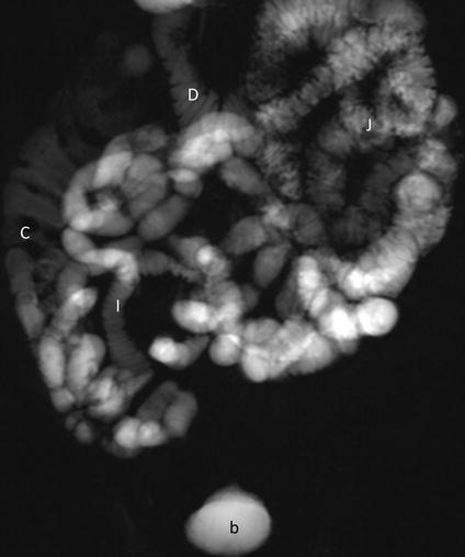
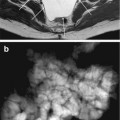
Fig. 3.1
MR fluoroscopy. A panoramic view of the small bowel. D duodenum, J jejunum, I ileum, C colon, b bladder
On MR images, the duodenal “C-shaped” (Fig. 3.2) as well as the duodenal and jejunal wall, lumen, and fold patterns are well demonstrated (Figs. 3.3 and 3.4). The bowel diameter, the fold thickness, and the number and interfold distance gradually decrease in size, reaching their smallest measurements in the terminal ileum (Figs. 3.1 and 3.5). Furthermore, the diameters of proximal, distal, and terminal ileum; its fold number; fold thickness; and interfold distances are not markedly dissimilar. The small bowel wall thickness is similar in size throughout the bowel measuring approximately 1.5 ± 0.5 mm [1].
Get Clinical Tree app for offline access
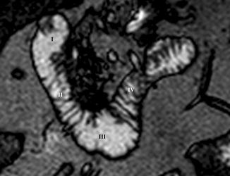
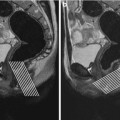
Fig. 3.2
Coronal TrueFISP image. Different parts of duodenum, which appear distended and with its characteristic fold pattern. (I): Superior part; (II): descending part; (III): horizontal part; (IV): ascending part
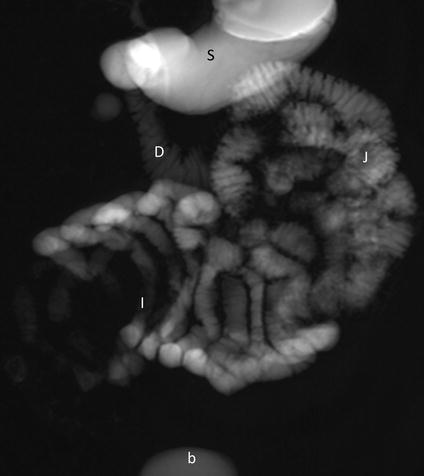
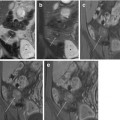
Fig. 3.3
MR fluoroscopy. Note how duodenum (D) and jejunum (J


Related posts:

Stay updated, free articles. Join our Telegram channel
Full access? Get Clinical Tree
