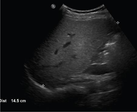
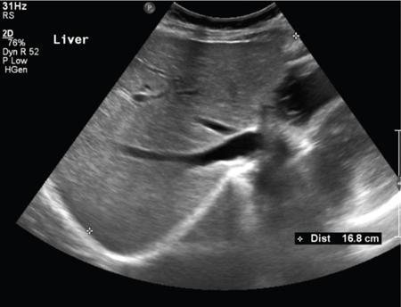
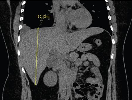
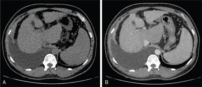
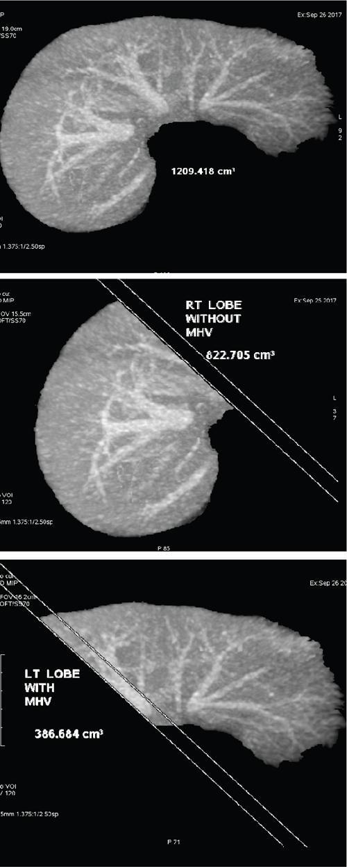
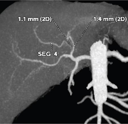
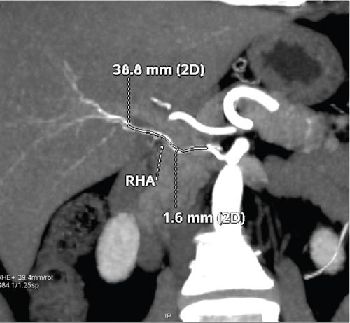
Ritu K. Kashikar, Shrinivas B. Desai Knowledge regarding normal dimensions of organs is important as visceromegaly is the first and often only abnormality in a variety of disorders. The radiologist should also be aware of normal diameters of vessels and ducts because an increase in size is usually a pointer to pathology in the organ. This chapter is a lucid review of normal sizes of organs, vessels, duct and also focuses on which section and location should size be measured to avoid interobserver variation. The liver is the largest organ in the abdomen. Hepatomegaly is a common condition and often the first clinical and imaging feature of various disorders. It is hence imperative for the radiologist to know the normal size and the section on which measurements should be taken. USG is commonly used to measure liver size. The longitudinal view is commonly used to measure liver size. The liver is considered normal in size if on longitudinal scan through the midhepatic line the liver measures 13 cm or less. This is true in approximately 93% individuals Measurement more than 15.5 cm suggests hepatomegaly in 75% cases (Figs. 9.3.1 and 9.3.2) (Table 9.3.1). Hepatomegaly is also suggested by an inferior angle of more than 45 degrees in the left lobe and more than 90 degrees in the right lobe. The normal liver measures approximately 6.5 cm first 3 months of age and reaches a size of 12.5 cm by 10–12 years of age. Liver size can be measured on unenhanced or enhanced CT. On CT the liver measures 10–12.5 cm in the midclavicular line on an average. A liver measuring more than 15.5–16 cm in the midclavicular line is considered enlarged. The midclavicular line measurement is done in coronal plane (Fig. 9.3.3). Another important measurement is the size of caudate lobe. The caudate to right lobe ratio (C/RL) is a measurement used to diagnose caudate lobe hypertrophy and right lobe atrophy which is important in the diagnosis of cirrhosis. The axial section immediately below main portal vein bifurcation is used for measurements. The following lines are drawn on the liver (Fig. 9.3.4). C/RL: In an adult patient of average weight (60 kg), the estimated liver volumes can range from 1024–1302 cm3 (Fig. 9.3.6). USG and Doppler provide important information regarding patency of artery in postoperative/transplant setting. Normal hepatic artery waveform is pulsatile and of low resistance. The normal resistive index measures 0.7. High or low resistivity index (RI) indicated pathology. The measurements of the hepatic arteries bare importance in transplant imaging. The diameter and length of the arteries are best measured on CT angiogram images. Arteries smaller in calibre than 2 mm may be difficult to anatomize. Replaced RHA is often longer in length than standard arteries. The normal diameters of the hepatic arteries are mentioned in Table 9.3.2 (Figs. 9.3.7–9.3.9). The portal venous system is valveless and hence its diameter is influenced by respiratory variations. The portal venous diameter is greatest during inspiration and hence all measurement should be made in this phase (Table 9.3.3). The diameter of portal vein has importance in diagnosing portal hypertension and USG is often used for this purpose. USG also provides other important parameters like flow velocity and volume flow which are relevant in the setting of portal hypertension. The normal portal venous velocity measures 15–18 cm/sec.(Fig. 9.3.10)
9.3: Normogram and normal values
Introduction
Liver
Normal usg measurements
Midclavicular line
The normal liver measures 10.5 ± 1.5 cm in longitudinal diameter and 8.1 ± 1.9 cm in the anteroposterior projection
Midline
Normal liver measures 8.3 ± 1.7 cm (95th percentile = 10.9 cm) and 5.7 ± 1.5 cm (95th percentile = 8.2 cm) in longitudinal and anteroposterior dimensions
Normal liver size by age
Measurement of liver size on CT
Measurement of caudate lobe
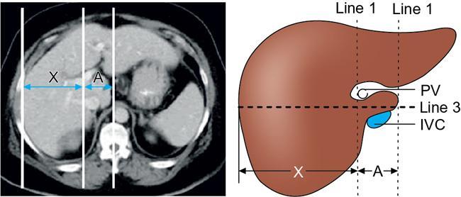
Interpretation
Normal hepatic volume
Hepatic artery
Common hepatic artery
0.50 ± 0.04 cm
Hepatic artery proper
0.45 ± 0.03 cm
Left hepatic artery
0.30 ± 0.03 cm
Right hepatic artery
0.36 ± 0.04 cm

Portal vein
Radiology Key
Fastest Radiology Insight Engine


