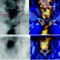
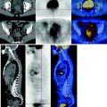
Fig. 73.1
PET scan showed abnormal glucose consumption of the right lung hilar nodule
Softer carbohydrate consumption of the right posterolateral tenth rib, SUV max 2.8.
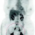
No areas of abnormal metabolism of the remaining parts of the body examined.
73.4 Conclusions
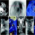
The PET scan shows poor response to treatment of lung cancer and bone metastases already mentioned in history. See Figs. 73.2, 73.3, 73.4.
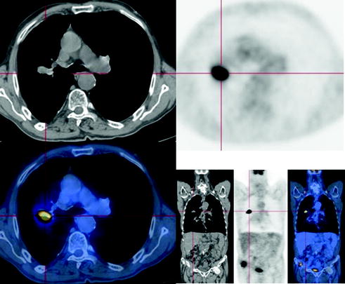

Fig. 73.2


In correspondence of the middle lobe of the right lung, CT-PET shows a solid nodule, uneven, with irregular margins and striae that connect it with the hilum. This mass is characterized by high glucose metabolism
Stay updated, free articles. Join our Telegram channel

Full access? Get Clinical Tree


