16 Nuclear medicine
Definition of nuclear medicine
| The introduction of a specific pharmaceutical (depending on which part of the body is to be targeted), which has been labelled with a radioisotope, into a patient. The gamma rays emitted by the radioisotope are scanned by a detector and the diagnostic image is produced showing the concentrations of the radiopharmaceutical (e.g. a bone scan) or an indication of function (e.g. the glomerular filtration rate of the kidneys) |
| Charge Collection | The pooling of electrons across a crystal |
| Gamma Camera | A large, stationary, scintillation counter, which records the activity over the whole field at the same time. Used to detect pathologies where the physiology of the structure is changed |
| Gantry | A structure or support, in which the X-ray tube, detectors and associated electronics are housed |
| Half-life | The amount of time taken for the radioactivity of a radioactive substance to decay by half the initial value. The half-life is a constant for each radioactive isotope |
| Image Fusion | When a PET (or SPECT) image and a CT image are viewed together by one being superimposed on the other |
| Pharmaceutical | A drug used in medicine |
| Photodiode | A semiconductor used to detect light and then generate electricity in proportion to the quantity of light detected |
| Photomultiplier | Equipment that produces an amplified current when exposed to electromagnetic radiation (light). Photons hitting the cathode produce electrons which in turn hit other surfaces thus producing more electrons, forming a pulse of electricity which forms the subsequent image |
| Planar | A two dimensional image |
| Pulse Height Analyser | Receives the signal from the photomultiplier and only produces an electrical signal if the input pulse lies in a predetermined range |
| Radioisotope | Any isotope that is radioactive. Forms of an element which have the same atomic number but different mass numbers, exhibiting the property of spontaneous nuclear disintegration |
| Radiopharmaceutical | A drug consisting of a radioactive compound |
| Scintillation Counter | A number of scintillator crystals in containers, one surface of the crystal is attached to a transparent glass window and the other surfaces are coated with magnesium oxide to reflect light back into the crystal; the back of the crystal is attached to a photomultiplier tube. If a gamma ray hits the crystal, light is produced and some reaches the photocathode of the photomultiplier |
| Scintillator | A sodium iodide (or caesium iodide) crystal with a thallium activator |
| Segmented | Divided into sections |
| Solid State Detector Material | Semi conductor – Cadmium zinc telluride (CdZnTe) |
| Spatial Resolution | The smallest distance between two objects that can be visually seen on an imaging system |
| Pharmaceutical | A drug which is absorbed by a specific (targeted) area of the body |
| Radioisotope | Technetium 99m (99Tcm) most commonly used |
• Is a gamma emitter
• Has a half-life of 6.02 hours
• Is readily available
• Is readily combined with pharmaceuticals
• Has lower emissions than other types of radiation
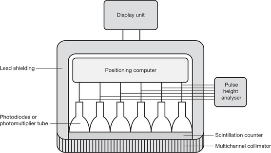
It is combined with a specific pharmaceutical so that a specific area of the body will take up the radioisotopeDetector – Gamma CameraRadiation from the patient passes through:
A multichannel collimator
• Parallel lead columns
• Absorb extraneous radiation
• Only allows gamma rays that are at 90° to the crystal face to reach the crystal
A scintillation counter
• Gamma rays from the patient hit the glass window
• Light is produced by the crystal
Photodiodes or photomultipliers
• Detects the light from the crystal
• Converts the light to an electrical pulse
Pulse height analyser
• Filters the electrical pulses
• Only allows pulses of a predetermined strength to be measured over time
• The signal then goes to the computer monitor
Fig. 16.1 The main parts of a gamma camera.
Operator ConsoleWhere the operator can determine the settings for the scanDisplay StationFor the viewing, analysis, networking and storage of the final image
| Rectilinear Scanners |
• Original scanner
|
Stay updated, free articles. Join our Telegram channel

Full access? Get Clinical Tree


