| SKULL BASE REGION | Sella turcica |
| HISTOPATHOLOGY | Null-cell pituitary adenoma |
| PRIOR SURGICAL RESECTION | Yes |
| PERTINENT LABORATORY FINDINGS | Prolactin: 49 ng/mL |
Case description
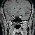
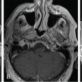
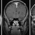
A 28-year-old woman with temporal visual field deficits and persistent galactorrhea after pregnancy was found to have a large hemorrhagic sellar lesion exhibiting a mass effect on the optic chiasm and hypothalamus with extension into the suprasellar region ( Figure 3.12.1 ). The patient underwent gross-total resection via an endonasal transsphenoidal approach ( Figure 3.12.2 ). Pathology revealed a pituitary adenoma, negative for all hormones. A 2-month postoperative follow-up with her ophthalmologist showed resolution of the visual field deficits.


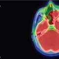
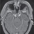
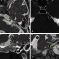
The patient was followed with annual imaging for 4 years with no progression of the tumor. She was lost to follow-up for another 3 years and, upon reestablishing care, she again complained of headaches and blurry vision. MRI revealed recurrence of the adenoma, with two nodules: one on the right side of the normal pituitary gland (in contact with the right optic nerve) and one along the central floor of the sella, extending to the midline ( Figure 3.12.3 ). Considering that the recurrent nonfunctioning pituitary adenoma was in immediate contact with the right optic nerve, the decision was made to pursue hypofractionated stereotactic radiosurgery (SRS) using the Gamma Knife radiosurgery (GKRS) machine ( Figure 3.12.4 ).
| Radiosurgery Machine | Gamma Knife |
| Radiosurgery Dose (Gy) | 25, at 50% isodose line |
| Number of Fractions | 5 |


