| CRANIAL REGION | Superior sagittal sinus |
| HISTOPATHOLOGY | Meningothelial meningioma, WHO grade 1 |
| PRIOR SURGICAL RESECTION | Yes |
| PERTINENT LABORATORY FINDINGS | N/A |
Case description
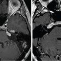
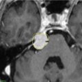
A 49-year-old female presented with headaches, speech disorder, and nausea. Magnetic resonance imaging (MRI) demonstrated an 80-cc left frontal parasagittal meningioma ( Figure 12.58.1 ). She underwent initial subtotal resection, and histopathology revealed a meningothelial WHO grade 1 meningioma. Two years after resection, she presented with headaches and short-term memory loss. Follow-up MRI showed tumor progression in the anterior sagittal sinus region, which was treated with Gamma Knife radiosurgery (GKRS) ( Figure 12.58.2 ). The treated tumor remained stable on follow-up. However, tumor progression was noticed outside the treated area in the falx and posterior superior sagittal sinus (SSS) at 18 months and 3 years from surgical resection, respectively. The tumor progression was then managed with three additional radiosurgical procedures (see Figure 12.58.2 ). Due to further tumor progression 6 years later, the patient underwent fractionated intensity-modulated radiation therapy (IMRT) (54 Gy, 30 fractions) as well as a fourth GKRS session. At 18 years after the craniotomy, the patient remained neurologically stable, without any neurological signs or symptoms.
| Radiosurgery Machine | Gamma Knife – Model C |
| Radiosurgery Dose (Gy) |
|
| Number of Fractions |
|

Related posts:
 Esthesioneuroblastoma – delayed postoperative radiosurgery for recurrence at long-term
 Null cell – delayed postoperative radiosurgery for growing perioptic residual
 Chondrosarcoma – definitive radiosurgery after subtotal resections
 Large vestibular schwannoma – delayed postoperative radiosurgery for growing residual
 Trigeminal neuralgia due to petroclival meningioma – upfront radiosurgery
 Superior sagittal sinus meningioma – immediate postoperative radiosurgery for residual
Stay updated, free articles. Join our Telegram channel

Full access? Get Clinical Tree


