| SKULL BASE REGION | Petroclival |
| HISTOPATHOLOGY | N/A |
| PRIOR SURGICAL RESECTION | No |
| PERTINENT LABORATORY FINDINGS | N/A |
Case description
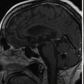
The patient presented with rapid onset of neuropathic right facial pain, which persisted for 6 months. The pain was partially relieved by pregabalin, but no neurologic deficit was otherwise present. Brain magnetic resonance imaging (MRI) revealed a petroclival meningioma with mass effect on the brainstem and right trigeminal nerve ( Figure 9.45.1 ). Due to the patient’s advanced age and prior medical history, stereotactic radiosurgery (SRS) was recommended to control tumor growth ( Figure 9.45.2 ).
| Radiosurgery Machine | Gamma Knife – Perfexion |
| Radiosurgery Dose (Gy) | 12 at the 50% isodose line |
| Number of Fractions | 1 |

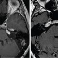
Postcontrast T1-weighted image (left) showing a lesion highly suspicious of meningioma in the petroclival region. A CISS sequence image (right) showing the tumor with mass effect on the brainstem. CISS, Constructive interference in steady state.

Imaging of the treatment plan.
| Critical Structure | Dose Tolerance |
|---|---|
| Brainstem | <0.01 cc >15 Gy |
| Cranial nerves in cavernous sinus | Unknown, but significantly more resistant than the optic nerve |
| Optic pathways | <0.01 cc >8 Gy |
| Modiolus | Maximum dose ≤4 Gy |
Related posts:
 Esthesioneuroblastoma – delayed postoperative radiosurgery for recurrence at long-term
 Null cell – delayed postoperative radiosurgery for growing perioptic residual
 Suprasellar non-small cell lung carcinoma metastasis – upfront radiosurgery
 Chondrosarcoma – definitive radiosurgery after subtotal resections
 Large vestibular schwannoma – delayed postoperative radiosurgery for growing residual
 Capillary hemangioma – postoperative radiosurgery for residual tumor
Stay updated, free articles. Join our Telegram channel

Full access? Get Clinical Tree


