| CRANIAL REGION | Superior sagittal sinus |
| HISTOPATHOLOGY | Meningothelial meningioma, WHO grade 1 |
| PRIOR SURGICAL RESECTION | Yes |
| PERTINENT LABORATORY FINDINGS | N/A |
Case description
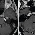
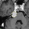
A 45-year-old female presented with a headache, slurred speech, and difficulty understanding speech. A subsequent magnetic resonance imaging (MRI) scan revealed a right parasagittal meningioma with skull invasion and extension into the superior sagittal sinus (SSS). She underwent subtotal resection ( Figure 12.59.1 ), followed by Gamma Knife radiosurgery (GKRS) ( Figure 12.59.2 ). Histopathology confirmed the diagnosis of a WHO grade 1 meningioma. After GRKS, the patient was maintained on Trileptal (300 mg TID) to control persistent seizures.
| Radiosurgery Machine | Gamma Knife – Model C |
| Radiosurgery Dose (Gy) | 13, at the 50% isodose line |
| Number of Fractions | 1 |

Axial, coronal, and sagittal postcontrast T1-weighted images showing a parasagittal meningioma (30 cc volume) invading the superior sagittal sinus bilaterally. The patient underwent a craniotomy for subtotal resection and cranioplasty using a mesh, and histopathology confirmed a diagnosis of WHO grade 1 meningioma.
Related posts:
 Esthesioneuroblastoma – delayed postoperative radiosurgery for recurrence at long-term
 Null cell – delayed postoperative radiosurgery for growing perioptic residual
 Chondrosarcoma – definitive radiosurgery after subtotal resections
 Large vestibular schwannoma – delayed postoperative radiosurgery for growing residual
 Trigeminal neuralgia due to petroclival meningioma – upfront radiosurgery
 Superior sagittal sinus meningioma – delayed postoperative, multisession radiosurgery for growing residual
Stay updated, free articles. Join our Telegram channel

Full access? Get Clinical Tree


