Fig. 1
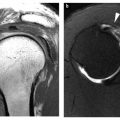
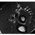
Preoperative staging. Multiple retroperitoneal lymph node metastases in a 66-year-old patient with prostate-specific antigen 88.4 ng/mL, Gleason score 8 (for color reproduction see p 311)
Bone Metastases
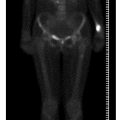
In the largest pre-operative series reported, our group found bone metastases in 13/130 patients pre-operatively; 2 of the 13 patients had bone metastases that had not been detected previously by conventional imaging (Fig. 2) [3]. The results of the study showed that FCH PET/CT would result in a therapy change for 15% of patients overall, and an upstaging change in 20% of high-risk patients. In evaluating 70 men with PCa, our group noted sensitivity, specificity and accuracy of 79%, 97% and 84%, respectively, for FCH PET/CT compared with a consensus definition of bone metastases based on conventional imaging and clinical endpoints [8]. In another prospective series of 38 patients with high-risk PCa [9], we compared the performance of FCH PET/CT with that of [18F]fluoride PET/CT (Table 1). FCH PET/CT showed significantly higher specificity than [18F]fluoride PET/CT and comparable sensitivity. However superior performance in the detection of bone marrow metastases was seen using FCH PET imaging. In those studies, discordant findings were noticed in a relatively small subset of patients with densely sclerotic lesions on CT that were negative in FCH PET and positive in [18F]fluoride PET scans (Fig. 3).
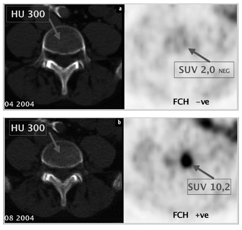
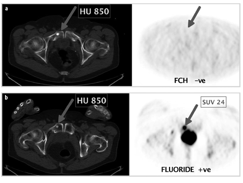

Fig. 2 a, b.
Preoperative staging. Bone marrow metastasis in L4 in a 73year-old patient with prostate-specific antigen 8.7 ng/ml, Gleason score 8. [18F]fluorocholine (FCH) PET/CT negative [standardized uptake value (SUV) 2.0] a, and positive after 4 months (SUV 10.2), b. HU Hounsfield units (for color reproduction see p 311)

Fig. 3 a. b.
Follow-up (prostate-specific antigen 20.5 ng/ml) 1 month after withdrawal of hormone therapy. Bone metastasis in the right os pubis [CT: sclerosis, Hounsfield units (HU) 850]. [18F]fluorocholine PET/CT negative (FCH –ve) (a) and [18F]fluoride PET/CT positive (FLUORIDE +ve) (b) [standardized uptake value (SUV) 24] (for color reproduction see p 312)
Table 1
Comparison of FCH PET/CT and [18F]fluoride PET/CT in 38 patients (preoperative n = 17, postoperative n = 21)
FCH PET – CT | |||
|---|---|---|---|
True | False | Total | |
Positive | 97 | 1 | 98 |
Negative | 95 | 34 | 129 |
Estimate value
Stay updated, free articles. Join our Telegram channel
Full access? Get Clinical Tree
 Get Clinical Tree app for offline access

| |||

