Fig. 1
Delayed whole body scintigram demonstrates the typical axial locations for metastatic disease
The location should be predominantly axial, and when in long bones mostly in the proximal metaphyses. Eighty percent of patients have axial involvement, 50% in the spine, and less commonly in the cervical spine. Thirty percent have lesions in the pelvis, 20% in ribs and 15– 20% in the skull. Long bone involvement is also seen in 15% of patients.
If one sees more distal metastases as well as proximal metastases, one should have a higher suspicion for an anemia causing redistribution of red marrow more distally. Distal lesions can also be seen in lung and, less commonly, breast cancer.
When presented with an image where there is a single isolated area that might be a metastatic deposit, our recommendation is to do another more sensitive examination to see if there are multiple deposits elsewhere. Only approximately 15% of these deposits are found to be metastases. The next most sensitive test will be positron emission tomography (PET) scanning most often or, in different centers, whole body MRI. In the majority of cases these focal deposits will be multifocal when a more sensitive test is performed.
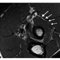
Diffuse osseous metastatic disease is termed skeletal carcinomatosis, and it is usually related to a breast or a prostate primary. On PET or skeletal scinitgraphy, a superscan is seen. The superscan is characterized by lack of renal uptake, more intense skull and metaphyseal activity, and less than usual soft tissue uptake (Fig. 2). Somewhat surprisingly, these patients can survive for a moderate amount of time.
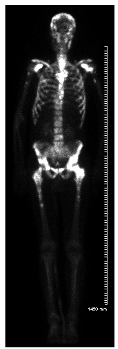
Fig. 2
Superscan in breast metastases with metaphyseal and skull increased uptake, and relative lack of renal and soft tissue uptake
Very rarely will metastatic deposits be cold on bone scan. Most often these are hypervascular, quite destructive lesions that yield little reactive bone formation in the natural history. The most commonly described primaries include renal cell cancer, occasional thyroid cancer and occasional colon cancer. On radiography, one will see a very lytic blown out lesion, sometimes with an accompanying soft tissue mass.
Follow-up of potentially treated metastases by scintigraphy is troublesome. It is easy when lesions are smaller or go away. However, increased activity might not represent worsening disease. On skeletal scanning, the flair phenomenon might be noted, with falsely increased activity in previously seen lesions. Usually no new locations are noted. However, very subtle lesions can heal and show the flair phenomenon and thus look like new lesions. This appearance usually reverts back to normal after about 3 months. Unfortunately, this is often too long to alter treatment, and other follow-up imaging, such as PET or MRI, is used.
PET Scanning
PET scanning should be rarely used in the initial evaluation of metastatic disease, not because it is not accurate, but because it is usually overkill. Sometimes when a PET scan is suggested, an MRI can be done at lower cost and with higher sensitivity. In addition, PET scanning is often less useful for less biologically aggressive metastases, such as some prostate metastases and breast metastases.
However, PET scanning is useful when there are indeterminate bone scintigram findings for metastatic disease or when a whole body PET scan is done for total body staging, including both the osseous and the nonosseous portions of anatomy. The distribution is as described above for scintigraphy, and characterized by similarly variable size, variable shape and variable intensity. When a single lesion is seen on PET scanning, a focal MRI of that area should be done. If the MRI is still nonspecific, then whole body marrow imaging should be performed. There are specific false-positive lesions on PET scanning that are more common than on scinitgraphy; thus closer radiologic correlation is often needed. This can usually be done via the concomitant CT. False-positive causes of PET scanning are protean, related to its high sensitivity and include arthroses, Paget’s disease, stress injuries and benign tumors.
F18 PET scanning is useful as a higher-sensitivity bone scan and gives a similar appearance for osteoblastic disease.
Related posts:
 Imaging of Bone Metastases with FDG, Fluoride and Gallium-Somatostatin Analogs
How to Interpret Asymmetries, Masses and Architectural Distortions
Imaging of Bone Metastases with FDG, Fluoride and Gallium-Somatostatin Analogs
How to Interpret Asymmetries, Masses and Architectural Distortions
 Rotator Cuff and Impingement Syndromes
 of the Upper Extremity
 of the Breast: An Approach to Radiologic Classification
 Imaging with an Emphasis on Magnetic Resonance Imaging
Stay updated, free articles. Join our Telegram channel

Full access? Get Clinical Tree


