Opening Round

Opening Round


Case 1


Comment


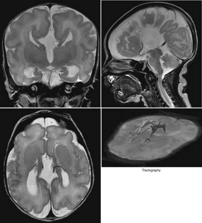
The CC is the largest commissure (bundle of white matter tracts) connecting both cerebral hemispheres. Additional hemispheric connections are the anterior commissure and the hippocampal commissure. The CC has a complex, programmed anterior-to-posterior development starting with the genu and followed by the truncus and splenium. The rostrum of the CC is the final segment to develop. Agenesis of the CC is observed on imaging with multiple, characteristic anatomic sequelae. Most of the classical sequelae are demonstrated in this case. The lack of the CC is usually evident in the midline, sagittal slice. In addition, the sulci along the medial surface of both cerebral hemispheres show a typical radiating appearance converging to the third ventricle. The third ventricle may be enlarged and extend interhemispherically. In rare cases the third ventricle may reach the vertex, or an associated interhemispheric cyst may be revealed. On axial imaging the lateral ventricles reveal a parallel course because the CC is lacking. In addition, frequently the occipital horns of the ventricles are enlarged (colpocephaly). The fibers that cannot cross the midline usually realign along the medial contour of the lateral ventricles and run in an anterior-to-posterior direction. These fibers are known as Probst bundles and can easily be recognized on tractography reconstructions using diffusion tensor imaging data. On coronal imaging the combination of the separated lateral ventricles, the medial impression of these ventricles by the Probst bundles, and the shape of the adjacent third ventricle mimic a trident or Texas longhorn cow. Because the CC is one part of the commissures connecting both hemispheres, the remainder of the commissures should be studied for additional malformations. The hippocampi may be malrotated; the anterior commissure may be lacking. In 50% of children a CC agenesis is part of a more extensive malformation (e.g., Dandy-Walker malformation, Arnold-Chiari II malformation, septooptic dysplasia). In addition, migrational abnormalities are frequently encountered. Ruling out additional malformations is essential; doing so will determine a functional and cognitive prognosis. Clinically, an isolated CC agenesis may be an incidental finding on an MRI. If additional malformations are present, then seizures, a developmental delay, and a hypothalamic-pituitary dysfunction may result. CC agenesis should be differentiated from secondary injury of the CC. For example, a severe atrophy of the CC resulting from an extensive periventricular leukomalacia should not be confused with a primary CC agenesis. In addition, it is important to remember that the only exception to the anterior-to-posterior rule of development is a semilobar or lobar holoprosencephaly. In these malformations the posterior CC may be present without the genu or anterior trunk of the CC.


Case 2


Case 3


Comment


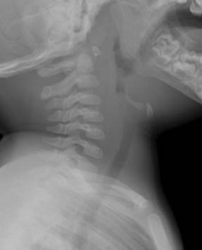
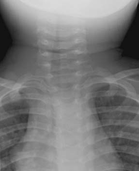
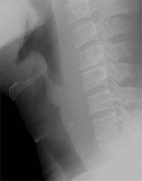
Common infectious causes of upper respiratory symptoms in childhood include epiglottitis, croup and/or tracheitis, and retropharyngeal abscess. Epiglottitis is usually caused by Haemophilus influenzae type B (HIB), which is much less common now that immunization against HIB is routine. Differentiation from the other causes of upper respiratory infection in part is determined by the clinical setting. Croup is often seen in a younger patient, 6 months to 3 years old; it is a viral illness and fever is usually not as high as in epiglottitis. Tracheitis is less common and has more extensive involvement of the trachea compared with croup. It is usually of bacterial origin and is seen in the same age group as croup. Retropharyngeal abscess may be a complication of bacterial tonsillitis, and clinical findings of tonsillar infection may be present. This illness can be seen in infants younger than 6 months old.


If imaging is needed, anteroposterior (AP) and lateral views of the neck are necessary. The AP view is most important in the identification of the subglottic narrowing seen in croup. If findings point toward retropharyngeal abscess, contrast-enhanced computerized tomography is usually performed.


In 25% of patients with epiglottitis, subglottic narrowing consistent with edema is also present. Causes of epiglottic enlargement other than infection are angioneurotic edema, aryepiglottic or epiglottic cyst, hemophilia (hemorrhage), and thermal injury sometimes seen in child abuse.


Case 4


Comment


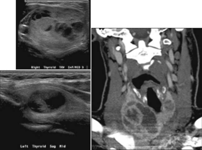
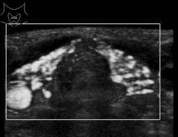
A goiter is a clinically recognizable enlargement of the thyroid gland that is often nodular. Goiters are endemic in areas of the world with iodine-deficient diets but also occur sporadically. Both endemic and sporadic goiters develop because of genetic predisposition and environmental factors such as iodine intake and cigarette smoking.


Most patients with nodular goiter are euthyroid, but they may become hyperthyroid or, less commonly, hypothyroid. This dysfunction occurs after the goiter has been present for many years. Clinically unapparent hyperthyroidism with low serum thyroid-stimulating hormone (TSH) and normal T4 and T3 is often present. As the TSH decreases, the size and nodularity of the gland may increase.


Ultrasound evaluation of the enlarged thyroid allows the detection and characterization of nodules, as well as guidance for fine needle aspiration and cytologic evaluation. Widespread use of this modality is due to its ready availability, low cost, minimal discomfort to the patient, and nonionizing nature. Cystic lesions are usually benign. When solid in appearance, lesions that are hypoechogenic have microcalcification and increased vascular flow; they are more likely to be malignant. Fine needle aspiration is the most direct technique to determine the makeup of a nodule and is widely used. I-123 nuclear studies are less sensitive to small nodules but in larger nodules can differentiate cold versus hot nodules (i.e., those that are not functioning versus those that have radioisotope uptake). Those nodules that function are highly unlikely to be malignant. In practice, this examination is not widely used in the setting of goiter evaluation. Computerized tomography and magnetic resonance image examinations offer reliable volume measurement and show the goiter extent, as well as its effect on adjacent structures.


In the absence of a malignant component and with normal thyroid function, the determination to treat a patient with goiter is mostly based on the symptoms from its mass effect and the cosmetic concerns. Total surgical thyroidectomy is the most common treatment. Ablation of the gland with iodine (I-131) therapy and L-thyroxine (L-T4) administration are other options.


Case 5


Comment


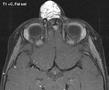
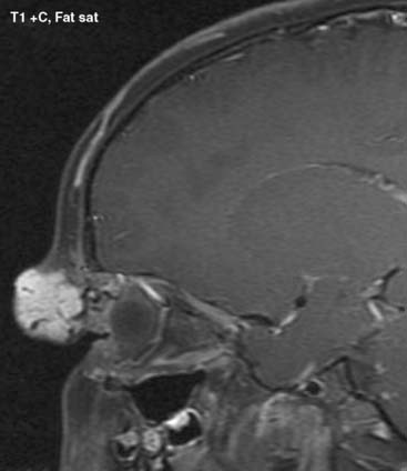
Hemangiomas are the most common tumors in infancy and childhood and account for 7% of benign soft tissue tumors. Hemangiomas are tumors that express a localized increase in angiogenic growth factors. Mulliken and Glowacki first described the classification of vascular anomalies based on clinical, histologic, and cytologic features. Vascular anomalies are divided into two major groups: (1) vascular tumors (hemangiomas), and (2) vascular malformations including venous malformations, lymphatic malformations, AVMs, and arteriovenous fistulas. The correct classification is important because treatment options differ significantly between the two groups. Hemangiomas have cellular proliferation of endothelial cells, and they are true neoplasms. Hemangiomas have subgroups consisting of infantile, congenital, noninvoluting, intramuscular, and kaposiform hemangioendothelioma types. The most common type is infantile hemangioma. Hemangiomas test positive for immunological markers such as glut1, FcrII, merosin, and Lewis Y antigen. A 3:1 female-to-male ratio exists.


Hemangiomas usually appear in the first week of life and can be located in the head and neck (60%), the trunk (25%), and the extremities. The patient displays a typical history of rapid neonatal growth (3 to 9 months) and slow involution characterized by hypercellularity during the proliferating phase, as well as fibrosis and diminished cellularity during the involuting phase (18 months to 10 years). This typical clinical history is oftentimes diagnostic; however, certain cases would require imaging confirmation.


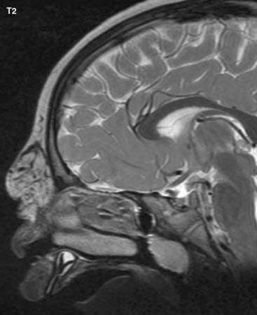
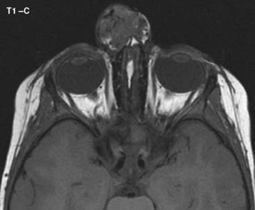
Imaging findings would obviously depend on the phase of the hemangioma. In the rapid-growth phase, ultrasound would reveal a variable echogenicity mass with increased flow on color-coded Doppler sonography. Involuting hemangiomas are heterogeneous masses with less intense color flow and fibrofatty changes. A magnetic resonance image (MRI) is advised with T1, T2, and postcontrast T1-weighted images. Fat suppression is of additional value in identifying this lesion. On MRI, typically a parenchymal, well-circumscribed mass with intermediate signal intensity on T1 and increased signal intensity on T2-weighted images is visualized. Avid contrast uptake is also noted, and the presence of flow voids within and around the soft tissue mass is an important feature.


In the majority of cases, no treatment is required because of spontaneous involution. Indications for treatment in the remainder of the cases are primarily functional, such as obscuration of vision or breathing or persistent cutaneous ulceration, high cardiac output failure. Systemic or intralesional steroid agents can be used for treatment. Promising results with systemic propranolol admission has been reported.


Case 6


Comment


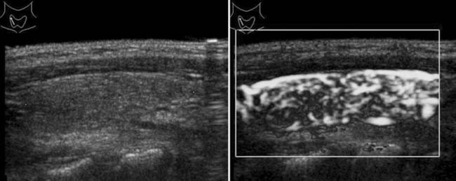
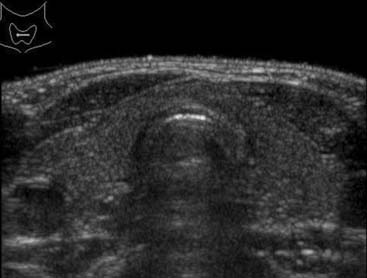
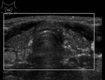
Hashimoto thyroiditis, also known as autoimmune thyroiditis or chronic lymphocytic thyroiditis, is the most common acquired disorder of the thyroid gland in children and the most common cause of hypothyroidism in the United States. Dr. Hakaru Hashimoto first described this disorder in 1912. Hashimoto thyroiditis is an autoimmune disease of unknown cause and may finally destroy the thyroid gland with resultant hypothyroidism. Incidence is estimated to be 1.3%. Blood tests are mandatory to determine the thyroid function by detecting the levels of thyroid hormones in general and to identify antithyroid peroxidase antibodies or antithyroglobulin antibodies in particular. Currently no cure exists for Hashimoto thyroiditis; however, the hypothyroidism can be treated effectively by substituting the deficient thyroid hormones. Symptoms are usually related to the progressive drop in thyroid hormones and not specific for Hashimoto thyroiditis. Hashimoto thyroiditis is more frequent in females and increases in frequency over age during childhood and adolescence.

Ultrasound examination is the primary imaging modality of choice. On ultrasound, the thyroid gland is usually diffusely enlarged, hypoechoic, and reveals significant hyperperfusion on color-coded Doppler sonography and power Doppler sonography, especially in the acute phase. Micronodulation characterized by multiple, small hypoechoic micronodules ranging between 1 and 6 mm in diameter are considered to be indicative of Hashimoto thyroiditis. Occasionally, discrete focal nodules are identified and fine needle aspiration cytology is necessary to confirm diagnosis. Hashimoto thyroiditis appears to occur more frequently in diabetic patients. Ultrasonography is also helpful to rule out additional lesions. Scintigraphy is rarely necessary to confirm diagnosis.


Case 7


Comment


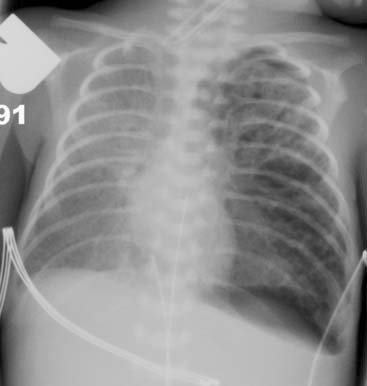
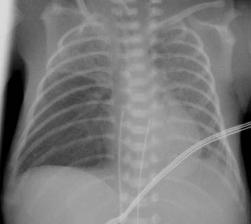
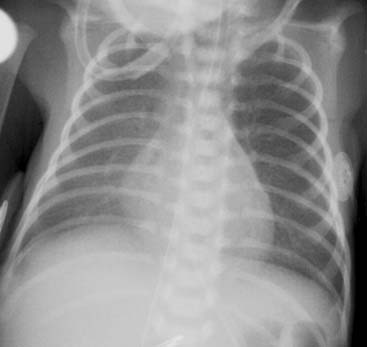
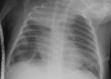
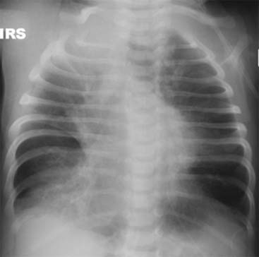
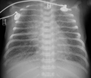
The lungs of a premature infant are stiff and noncompliant. This is due to lack of surfactant, normally produced by type II alveolar cells, beginning in week 24 of gestation and usually complete by week 32. Without this protein, alveolar distension and adequate oxygenation must be maintained by high-pressure ventilation with oxygen-rich air. This combination injures the already fragile alveolar walls and allows air to leak between the lining cells into the interstitial spaces and lymphatics. The air is first visible lying parallel to the bronchi, forming characteristic lucent lines and dots. Movement of the lungs with respiration causes the air to change configuration. It can dissect centrally or peripherally, causing pneumomediastinum and pneumothorax, or it can become centrally confluent, creating a pneumatocele. Air in the mediastinum can dissect downward through the inferior pulmonary ligaments and result in sterile pneumoperitoneum.


Aggressive treatment with exogenous surfactant in the delivery room can decrease the severity of the lung disease and avert this complication. Once present, it is treated by decreasing ventilatory pressures. Jet ventilation allows greater control of airway pressures. If unilateral, decubitus positioning onto the affected lung can effectively decrease pressure locally and hasten healing. Pneumatoceles occasionally must be surgically removed if they become intractably large.


Chest radiographs, which are a valuable monitor for this disease, can alert clinicians to dangerous levels of hyperinflation that could precipitate air leaks, enabling diagnosis of the emphysema and its complications and determining efficacy of therapeutic measures.


Case 8


Case 9


Case 10


Comments


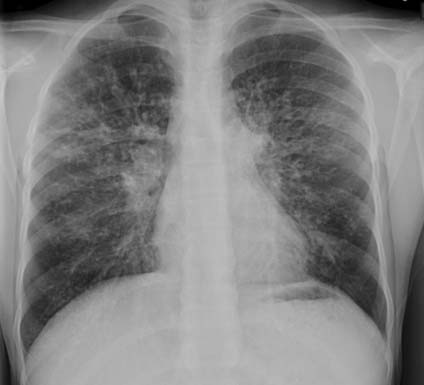
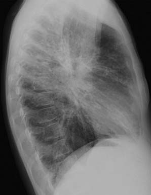
CF is an inherited, autosomal recessive disorder of exocrine gland function. The most common manifestations of CF are chronic respiratory infection and pancreatic enzyme insufficiency. CF is caused by defects in the gene for cystic fibrosis transmembrane conductance regulator (CFTR), which results in decreased secretions of chloride, increased reabsorption of sodium and water across epithelial cells, and ultimately to abnormal sticky mucous production. This viscous mucous is less effective in clearing secretions and results in a lung environment conducive to infection, as well as the other manifestations of this disease. CF is diagnosed by an abnormal sweat chloride test. The sweat is analyzed for its chloride content—the chloride will be high in CF. Specific gene typing is useful in further characterizing the disease, because more than 1400 types of mutations have been found in the CFTR gene.


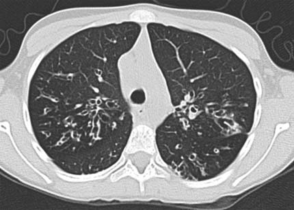
The radiographs shown here reveal the typical findings of hyperinflation, diffuse interstitial thickening, tram tracking of bronchiectasis, and mucous plugging. Although diffuse disease is usual, focal infiltrates and/or focal areas of more severe disease are common. As in this case, hilar enlargement because of lymphadenopathy (a manifestation of chronic infection) often is seen. Computed tomography (CT) examination generally shows more involvement than evident on chest radiographs. CT findings include, but are not limited to, peribronchial thickening, centrilobular opacity, bronchiectasis, mucous plugging, hilar adenopathy, bullae formation, and lung abscess.


The radiographic findings are nearly pathognomic for CF; hence the use of the term Aunt Minnie in this case. Rarely, other causes of chronic inflammatory disease may resemble CR; these include asthma and immunodeficiency syndromes.


Hemoptysis is common in advanced cases, and arteriography and direct embolization of the bronchial arteries may be necessary to treat it.


Case 11


Comments


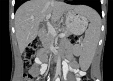
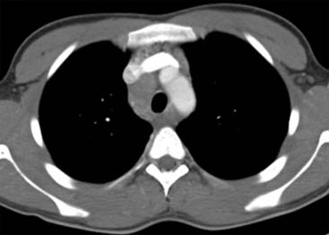
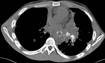
This 16-year-old boy, whose images are shown in this case, presented with a several-month history of fever, night sweats, fatigue, bone pain, and weight loss. He was found to have pancytopenia. Lymph node biopsy showed classical Hodgkin lymphoma, and he was staged as IV B because of apparent bone marrow involvement seen on PET scanning (bone scan and bone radiographs were negative).


Hodgkin disease is a common hematological malignancy that has two peaks of incidence: (1) between 15 and 35 years and (2) older than 55 years. The commonly used staging system (cited previously) is the Ann Arbor Staging System. In addition to the areas of involvement, bulky mediastinal disease involving more than one third of the intrathoracic diameter is also taken into consideration. Varying definitions of low, intermediate, and high-risk groups include all of these factors, as well as the histology.


The World Health Organization classification separates Hodgkin lymphoma into two groups: (1) classical (which includes lymphocyte-depleted, nodular-sclerosing, mixed cellularity, and classical lymphocyte-rich cell types) and (2) the lymphocyte-predominant type. Ninety percent of Hodgkin lymphoma is of the classical type, with Reed-Sternberg cells, and positive for CD15 and CD30 (CD stands for cluster of differentiation and is a protocol used to identify and investigate the cell surface molecules present on leukocytes). Immunohistological differences are seen between the subgroups of classical Hodgkin lymphoma, but the response to treatment is similar. Lymphocyte-predominant Hodgkin lymphoma expresses markers not usually seen in the classical type; those are B cell markers (CD20, CD79a, CD75) and epithelial membrane antigen and lymphocyte marker (CD45). This type of Hodgkin disease has a more indolent course and good prognosis with treatment, but the patient has a slightly higher risk of developing non-Hodgkin lymphoma.


Case 12


Comment


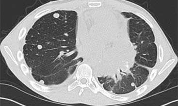
Nodular lung disease consists of multiple round opacities that can range from 1 mm to 1 cm or larger. The smaller lesions are only visible on computed tomography examination and are referred to as miliary. Further characterization of the nodules includes a description of their margins (smooth or irregular), presence or absence of cavitation, calcification, and their distribution.


Multiple lesions of different sizes, especially in subpleural or peripheral locations, suggest metastatic disease. Multiple small smooth or irregularly marginated nodules in a perilymphatic distribution suggest sarcoidosis. Silicosis and coal workers’ pneumoconiosis may also have this appearance. Upper lobe predominance is seen in coal workers’ pneumoconiosis. Small nodules of ground-glass opacity can be seen in extrinsic allergic alveolitis or bronchiolitis. Miliary or larger nodules are seen in the hematogenous spread of tuberculosis, fungal infection, or metastatic disease. If the lesions include some with thin-walled cavities, Langerhans cell histiocytosis should be considered. Other lesions that cavitate include metastatic squamous cell carcinoma, Wegener granulomatosis, rheumatoid lung disease, septic emboli, and multifocal infection. Lymphoproliferative disorders, lymphoma, leukemia, and Kaposi sarcoma may cause irregular nodules in a bronchovascular distribution. Calcification can be seen in granulomatous disease, hamartomas, metastatic tumor such as osteosarcoma, rarely as a consequence of infection and in abnormal calcium metabolism, with so-called metastatic pulmonary calcifications.


The patient in this example had osteosarcoma of the proximal femur with lung metastases at presentation 5 years earlier. By the time of these images, the patient had multiple tiny lesions (>100 total) resected from each lung. Chemotherapy is the initial treatment of nonmetastatic and metastatic osteosarcoma. Surgical resection of the primary lesion generally follows chemotherapy. Experimental administration of the bone-seeking radioisotope samarium (if the lesions are calcified and avid on nuclear bone scan) has recently been used to treat metastatic disease in osteosarcoma. The outcome in patients with lung metastases from osteosarcoma is improved by the combination of chemotherapy and surgery. A strong correlation exists between the degree of necrosis of the aggregate metastatic disease and the need for reoperation. In one study, those that needed reoperation had less than 80% necrosis of metastases.


Case 13


Comment


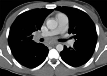
CT of the lung is used to detect infection in the immunocompromised patient. It is considerably more sensitive to early infection than chest radiography. In patients with febrile neutropenia, it is reported that a CT will find 20% more pneumonias 5 days earlier than the chest radiograph. In addition to increasing the detection rate, a careful analysis of the CT features can help to determine the likely cause of the infection.


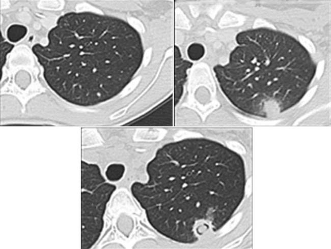
The patient in this example had invasive aspergillosis. The typical findings include single or multiple nodules, often with surrounding ground-glass opacity, the so-called halo sign. A lesion may cavitate and contain a solid nodule within it. This is called the air crescent sign. These features were all present in the patient.


The halo sign is seen in about half of patients with invasive aspergillosis and is caused by hemorrhage and necrosis around the necrotic nodule, which contains the fungal hyphae. This sign can also be seen in some bronchopneumonia, as well as tumors such as adenocarcinoma, alveolar cell carcinoma, Kaposi sarcoma, and metastasis. About 40% of nodules seen in fungal pneumonia are cavitary, and one half of them demonstrate the air crescent sign. This finding is not specific for invasive Aspergillus spp.; however, in the proper setting, it is highly suggestive for fungal disease (clinicians believe it to be caused by retracted and infarcted lung tissue within the cavity).


Although nodular disease is the most frequent manifestation of Aspergillus spp. infection on CT, ground-glass opacity and consolidation can also be seen. Other fungal infections are less common; increasingly pulmonary candidiasis is the cause of fever in the immunocompromised patient and its appearance is similar to Aspergillus spp.


Case 14


Case 15


Comment


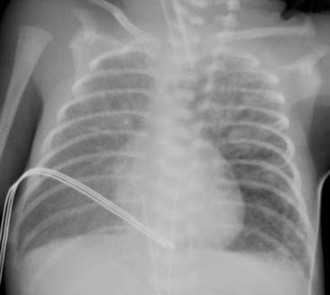
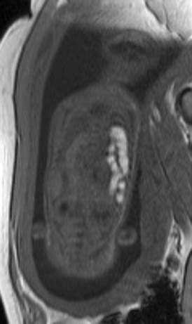
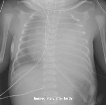
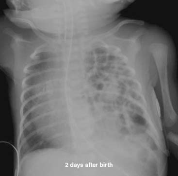
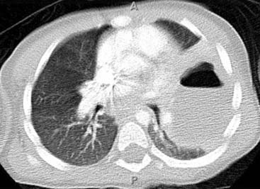
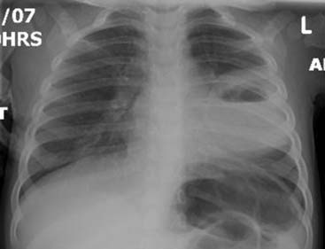
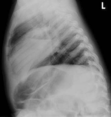
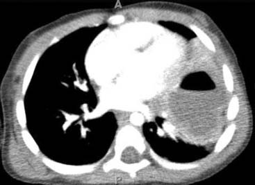
CDH occurs in 1 of every 2000 to 3000 live births and accounts for 8% of all major congenital anomalies. CDH can be described as cases in which abdominal contents are herniated into the chest. The three basic types of CDH include (1) the posterolateral Bochdalek hernia (occurring at approximately 6 weeks gestation), (2) the anterior Morgagni hernia, and (3) the hiatus hernia. The left-sided Bochdalek hernia occurs in approximately 85% of cases. Bochdalek hernia is more common on the left than on the right (5:1). The hernia may have variable abdominal contents: stomach, small and/or large bowel, liver, and spleen. Most common signs and symptoms include severe respiratory distress. This type of hernia typically presents at or soon after birth and is often detected at prenatal imaging. Less severe cases may present later in life or incidentally on radiograph. Large CDH results in pulmonary hypoplasia and pulmonary hypertension. Prognosis is primarily related to the degree of lung hypoplasia and pulmonary hypertension. Fetal magnetic resonance image (MRI) and fetal ultrasound have provided valuable information about lung volume, which is an important prognostic indicator in predicting pulmonary capacity after birth. Overall the survival rate is about 50%. Prenatal diagnosis, supportive care for pulmonary hypoplasia and respiratory failure, and use of extracorporeal membrane oxygenation (ECMO) resulted in improved survival rates. In utero surgical repair is an option when in utero diagnosis is made with ultrasound and MRI. Early diagnosis is crucial in guiding the mode of delivery (ex utero intrapartum treatment [EXIT] procedure) and immediate postnatal care. Surgical outcome is still variable. Up to one third of the cases have associated major malformations. Malrotation or stomach volvulus can be seen as a gastrointestinal tract abnormality, and 50% of the cases may have congenital heart disease.


Dec 21, 2015 | Posted by in PEDIATRIC IMAGING | Comments Off on Opening Round

Full access? Get Clinical Tree

Get Clinical Tree app for offline access