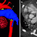
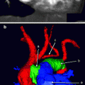
Fig. 11.1
Sagittal maximum-intensity projection (MIP; a) and color-coded three-dimensional (3D; b) images showing a Sano shunt (C) connecting the right ventricle (A) to the main pulmonary artery (D). Notice the stenotic portion of the proximal shunt (B), where the conduit traverses the right ventricular wall
Modified BT Shunt
The modified BT shunt uses a vascular graft (typically Gore-Tex [W. L. Gore and Associates, Elkton, MD]) that shunts blood from the subclavian or brachiocephalic artery to the ipsilateral main pulmonary artery. It often is used as a palliative procedure to increase pulmonary flow and expand the pulmonary arteries before more definitive surgery. The modified shunt differs from the original procedure, which is performed without a graft.
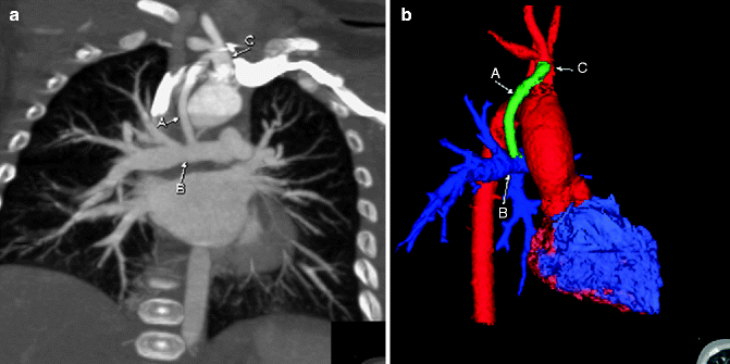

Fig. 11.2
Enhanced coronal (a) and color-coded 3D (b) images showing a modified BT shunt (A) connecting the right brachiocephalic artery (C) to the right main pulmonary artery (B)
Glenn Shunt
The Glenn shunt is a vascular conduit that shunts blood from the superior vena cava (SVC) to the right pulmonary artery. It is used in a staged palliative procedure in patients with single-ventricle physiology. Eventually, a more definitive Fontan shunt is performed to also supply blood from the inferior vena cava (IVC) to the pulmonary arteries. Glenn shunts typically are performed at 3–9 months of age, when pulmonary vascular resistance has decreased. If a left SVC also is present, bilateral bidirectional Glenn shunts are performed.
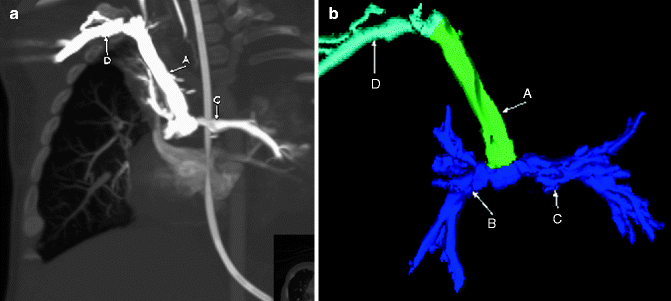

Fig. 11.3
Coronal MIP (a) and color-coded 3D (b) images showing a Glenn shunt channeling upper-extremity blood flow from the SVC (A) to the right pulmonary artery (B). The flow is bidirectional, with the left pulmonary artery (C) also filling. Contrast was injected from the right upper extremity, with the right subclavian vein (D) also opacified
Fontan Procedure
The Fontan procedure involves the placement of a vascular shunt that channels blood from the IVC to the pulmonary artery. It is the final procedure for patients with single-ventricle physiology to provide definitive blood flow to the pulmonary arteries. The surgery may be intra-atrial, in which a tunnel is created to channel blood to the pulmonary arteries, or extra-atrial in which a synthetic graft is placed to connect the IVC to the pulmonary arteries.
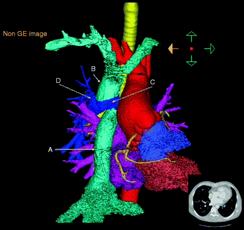

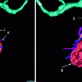
Fig. 11.4
Color-coded 3D image from a CTA scan showing an extracardiac Fontan graft (A) extending from the IVC to the main pulmonary artery (C). Also notice the presence of a Glenn shunt (B) channeling blood from the SVC to the right main pulmonary artery (D)
Hybrid Procedure
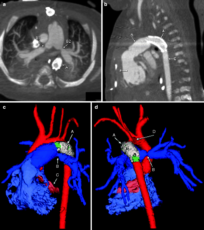
The hybrid procedure typically is performed for palliation in patients with hypoplastic left heart syndrome who may not be candidates for a stage I Norwood procedure. In the hybrid procedure, which does not require cardiopulmonary bypass, a vascular stent is placed in the patent ductus arteriosus (PDA) while bilateral pulmonary banding is performed to limit pulmonary blood flow.
