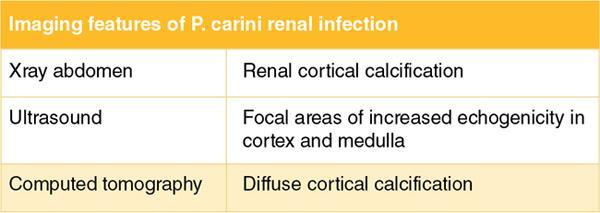
AIDS is an immunocompromised state and affected individuals are more prone for infections. The clinical course of AIDS is characterized by recurrent infections by uncommon opportunistic bacteria, fungi and viruses and they can involve any part of the urogenital system. Opportunistic organisms affecting kidneys in HIV-infected individuals include: This chapter mainly includes P. jiroveci and fungal infections. Tubercular infection is dealt with in a separate chapter. P. jiroveci infection is an AIDS defining illness, most commonly affecting the lungs and causes pneumonia. Extrapulmonary P. jiroveci infection occurs in only 1% of patients with P. jiroveci pneumonia. Risk factors of extrapulmonary infection in HIV-infected patients are low CD4+ count, history of other infection and aerosolized pentamidine prophylaxis (given for P. jiroveci pneumonia). Extrapulmonary infection can occur as a complication of P. jiroveci pneumonia. It can affect kidneys, liver, spleen, lymph nodes, adrenal glands, gut, bone marrow and brain. It spreads to kidney by the haematogenous and lymphatic routes from lungs. Renal involvement can be part of disseminated infection and can cause renal failure. Kidneys show multiple calcific nodules, especially in the cortex which represents areas of infiltrates and subsequent destruction of the renal tubules. With the use of silver stain. Pneumocystis jirovecii organisms will be evident. Azotemia, hypoalbuminemia, proteinuria and renal failure. Calcifications are common in pneumocystisis infection. On ultrasound, focal areas of increased echogenicity are seen in the cortex and medulla. On abdominal radiography, renal cortical calcifications are seen. Computed tomography shows diffuse cortical calcification. Presence of calcification and nephrocalcinosis are also seen in other renal infections such as M. avium-intracellulare or histoplasmosis and it is suggestive of HIV-associated infection but not specific for any pathogen. A summary of the various imaging features is shown in Fig. 10.12.2.10.1.
10. Opportunistic renal infections in HIV
Renal Pneumocystis jiroveci infection
Introduction
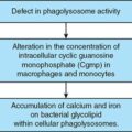
Etiopathogenesis
Histopathology
Clinical features
Imaging features
Related posts:


Stay updated, free articles. Join our Telegram channel

Full access? Get Clinical Tree


