Osteoporosis
Bone Densitometry
The basis for the radiologic diagnosis of osteoporosis is the measurement of bone mineral density (BMD). The standard clinical tests for this purpose are dual X-ray absorptiometry (DXA) and quantitative computed tomography (CT).
Dual X-Ray Absorptiometry
Dual X-ray absorptiometry has become established as the standard procedure for measuring BMD because of its high reproducibility, standardized methodology, and lower radiation exposure compared with quantitative CT.
In 1994 the World Health Organization (WHO) issued a definition for osteoporosis based entirely on bone densitometry by DXA. According to this definition, osteoporosis is present when the BMD is more than 2.5 standard deviations below the mean value for a healthy young reference population. The BMD expressed in relative standard deviations from the mean is called the T-score. Strictly speaking, the WHO definition applies only to density measurements performed in the lumbar spine (anteroposterior and posteroanterior projections) and proximal femur.
Bone mass is determined in relation to surface area and is expressed in g/cm2.
WHO definition of osteoporosis
• Normal: | T-score > – 1 |
• Osteopenia: | – 2.5 < T-score < – 1 |
• Osteoporosis: | T-score < – 2.5 |
Besides the T-score, which states the deviation of BMD from a healthy young reference population, DXA also provides a Z-score, which states the deviation from a reference population of the same age and gender.
The T-score is the most important parameter for predicting fracture risk and should direct treatment in consideration of other factors. The Z-score is of minor importance since it does not provide a direct measure of individual fracture risk.
Quantitative Computed Tomography
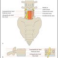
Bone densitometry by quantitative CT (QCT) is performed in the lumbar spine. The L1 through L3 vertebral bodies are scanned in transverse midvertebral sections (section thickness: 8–10 mm). A relatively large section thickness is used because averaging over a larger volume improves the reproducibility of the test. If the scout view shows the presence of a vertebral body fracture, an adjacent vertebra should be used for densitometry.
After the examination, a region of interest (ROI) is selected in the bone and the corresponding attenuation value is measured in Hounsfield units (HU). Conversion to an absolute density value is aided by using a reference phantom that is scanned with the patient. The result is expressed in mg/mL, i.e., bone mass relative to volume. The following cutoff values are used in the diagnosis of osteoporosis and osteopenia:
BMD values
Osteoporosis: < 80 mg/mL
Osteopenia: 80 mg/mL < BMD < 120 mg/mL
Related posts:

Stay updated, free articles. Join our Telegram channel

Full access? Get Clinical Tree


