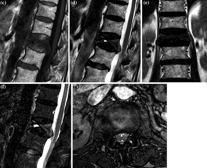
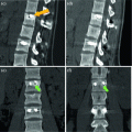
Fig. 1
a–g. XR (a), CT MPR sagittal (b), SE T1 sagittal (c), FSE T2 sagittal (d) and coronal (e) STIR sagittal (f), GE axial (g). L1 collapse, edema pattern (hyperintensity in f), intracanalar expansion of rear profiles with anterior epidural space obliteration, no compressive myelopathy
< div class='tao-gold-member'>
Only gold members can continue reading. Log In or Register to continue
Related posts:
 Herniated Lumbar Disk Diskectomy
 Dorsal Herniated Disk Diskectomy and Stabilization
 Lumbar Stenosis and Degenerative Instability Posterior Rigid Stabilization
 Traumatic Lumbar Collapse Rigid Stabilization
 Lumbar Collapse in Chordoma Vertebral Drawing
 Dorsal Collapse in Multiple Myeloma Vertebroplasty
Stay updated, free articles. Join our Telegram channel

Full access? Get Clinical Tree


