Other Gliomas
Overview
There are three rare gliomas—chordoid glioma, angiocentric glioma, and astroblastoma—that do not fall into a specific glioma category or classification. Each has unique molecular and genetic profiles, predilection for certain locations and ages, and imaging features.
|
Chordoid Glioma of the Third Ventricle
Definition: Chordoid glioma of the third ventricle is a low-grade, solid intraventricular glial neoplasm located in the third ventricle with indolent and noninvasive growth pattern and histologically composed of epithelioid cells expressing Glial fibrillary acidic protein (GFAP) in a background of mucinous stroma and lymphoplasmacytic infiltrate.
Epidemiology: This rare tumor occurs primarily in adults, with a female-to-male ratio of 2:1.
Affected age group: There is wide variation in age, with reported cases ranging from 5 to 71 years of age. Most patients are adults aged 35 to 60, with a mean age of 46 years.
Molecular and genetic profile: The most distinct immunohistologic feature is strong, diffuse positivity for glial fibrillary acidic protein as well as vimentin and CD34. Neuronal and neuroendocrine markers are absent. Chromosomal heterozygosity losses at 11q13 and 9p21 are noted.
CSF markers: None.
Clinical features and standard therapy: Surgical resection is the definitive therapy, and gross total resection usually results in cure. However, local recurrence can be seen in the setting subtotal resection.
Imaging
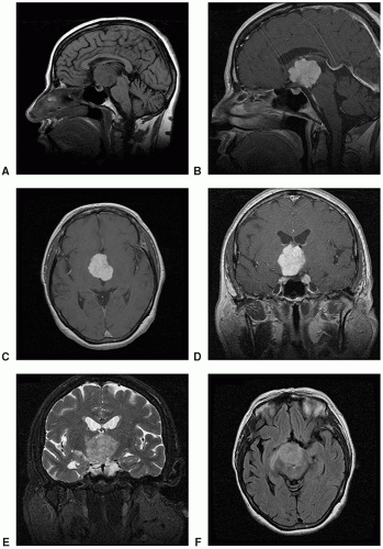
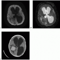
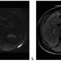
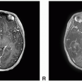
Chordoid glioma of the third ventricle is a solid, well-circumscribed tumor attached to the anterior wall or the roof of the third ventricle or inseparable from the hypothalamus.
Angiocentric Glioma
Definition: Angiocentric glioma is a circumscribed, superficially located, cortically based, benign cerebral tumor with angiocentric growth pattern and ependymal differentiation
Stay updated, free articles. Join our Telegram channel

Full access? Get Clinical Tree


