Ovarian cancer is the second-most common gynecologic malignancy and one of the leading causes of cancer-related deaths in women. However, its incidence remains low, accounting for approximately 1% of all types of cancers. Ovarian cancer encompasses a heterogeneous group of tumors classified based on distinctive histopathologic and molecular features. Epithelial ovarian tumors are the most common type, accounting for approximately 90% of ovarian cancers, and can be further divided into type I and type II cancers. Type II epithelial ovarian cancers are the most common and most aggressive, with high-grade serous carcinoma being the most common subtype.
Key points
-
•
Ovarian-Adnexal Reporting and Data System (O-RADS) ultrasound (US) and MRI are created to standardize reporting and communication of adnexal lesions; however, the focus of O-RADS is not screening for ovarian cancer but rather risk stratification of incidental adnexal lesions.
-
•
O-RADS US utilizes standardized lexicon terms to characterize ovarian/adnexal lesion, stratify the risk of malignancy, and provide a management system. However, individual case management may be modified, particularly in high-risk patients.
-
•
MRI is used as a secondary modality in O-RADS risk stratification and plays an important role in assessing sonographically indeterminate adnexal lesions and tumor subtyping. O-RADS MRI is applicable in average-risk patients with no acute symptoms. Ovarian/adnexal lesions in patients at high risk for ovarian cancer may have different positive predictive values.
-
•
QMRI are pivotal tools to differentiate benign from malignant lesions, with the ultimate goal of improving ovarian cancer outcomes and decreasing unnecessary surgeries for benign lesions.
Abbreviations
| AI | artificial intelligence |
| CfDNA | cell-free deoxyribonucleic acid |
| HGSC | high-grade serous carcinoma |
| O-RADS | Ovarian-Adnexal Reporting and Data System |
| PPV | positive predictive value |
| ROM | risk of malignancy |
| ROMA | Risk of Ovarian Malignancy Algorithm |
| STIC | serous tubal intraepithelial carcinoma |
| TIC | time intensity curve |
| US | ultrasound |
Introduction
Ovarian cancer is the second-most common gynecologic malignancy and one of the leading causes of cancer-related deaths in women. In 2025, there will be an estimated 20,890 new cases and 12,730 cancer-related deaths in the United States. However, its incidence remains low, accounting for approximately 1% of all types of cancers. Localized ovarian cancer has a relatively good prognosis with a 5 year survival of 92%; however, more than half of cases are detected at an advanced stage, with 5 year survival rate that drops to 72% if there is regional disease, and to 31% if there is distant disease. Current clinical trials and studies have not proven that ovarian cancer screening reduces mortality in average-risk asymptomatic patients, mainly due to its low incidence and lack of symptoms in early-stage disease. An effective approach to screen for ovarian cancer with adequate sensitivity and specificity in the general population remains an unmet clinical need and is the first focus of this article. On the other hand, ovarian and adnexal lesions are often incidentally detected on imaging performed for unrelated reasons, and radiologists are often faced with the task of risk stratification of these lesions. The goal in these situations is not to overlook potential malignancy while reducing unnecessary additional imaging or surgeries. Multiple imaging risk stratification strategies have been developed for these situations in recent years, particularly the Ovarian-Adnexal Reporting and Data System (O-RADS), which will be the second focus of this article.
Types of ovarian cancers
Ovarian cancer encompasses a heterogeneous group of tumors classified based on distinctive histopathologic and molecular features. , Primary ovarian cancer can be broadly divided into epithelial cell, germ cell, and sex cord-stromal tumors. Epithelial ovarian tumors are the most common type and account for approximately 90% of ovarian cancers. Epithelial ovarian cancers can be further subdivided into type I and type II, with distinct molecular profiles and clinical behaviors.
Type I epithelial ovarian cancers are lower-grade, more indolent, and less aggressive than type II epithelial ovarian cancers. These are often characterized by mutations in mitogen-activated protein kinase regulator pathways (eg, KRAS or BRAF). These include low-grade serous carcinoma, low-grade endometrioid carcinoma, clear cell carcinoma, and mucinous carcinoma. These tumors are more likely to be detected at an early stage by screening, given their indolent growth rate. In contrast, Type II epithelial ovarian cancers are the most common and most aggressive. These are associated with high mutation rates in TP53, somatic and germline mutations in BRCA1/2 and often have high genetic instability. These include high-grade serous carcinoma (HGSC, the most common subtype and often diagnosed at an advanced stage, Fig. 1 ), high-grade endometrioid carcinoma, carcinosarcoma, and undifferentiated carcinomas. The aggressive nature of high-grade disease limits the efficacy of ovarian cancer screening.
A 73 year old postmenopausal patient with an incidentally detected right adnexal mass found on a right hip MR imaging examination. Transvaginal pelvic ultrasound image with color Doppler of the right adnexa showed a 7 cm multilocular cystic mass with a solid component and a color score of 2. No ascites or peritoneal thickening was seen. This is an O-RADS US 4 lesion. Her CA 125 was 46 U/mL. Surgical pathology showed high-grade serous carcinoma of the right ovary with microscopic deposits involving the left ovarian surface (<1 mm) and omentum (<1 mm), consistent with stage IIIA disease.
Evidence has shown that the precursor for HGSC is serous tubal intraepithelial carcinoma (STIC) in the fallopian tube, rather than the ovary. This changing paradigm has significant implications for HGSC prevention (eg, opportunistic salpingectomy) and potentially screening. STIC is rare in patients undergoing salpingectomy for benign disease (<0.01%) but much higher in patients with a BRCA 1/2 pathologic variant (up to 2.8%). , The rate of HGSC after an isolated STIC diagnosis in patients undergoing prophylactic salpingo-oophorectomy has been reported to be around 7%, with a cumulative incidence of 10.5% after 5 years and 21.6% after 10 years. This observation has prompted further investigation into alternative approaches, including prophylactic salpingectomy with delayed oophorectomy, opportunistic salpingectomy, and salpingectomy alone. ,, Future imaging research focusing on the appearance of this precursor and early-stage disease in the fallopian tube could potentially help develop targeted screening and early detection of ovarian cancer.
Risk factors for ovarian cancer
Ovarian cancer incidence increases with age, with most ovarian cancer cases occurring after menopause, mainly affecting women aged 55 to 70 yrs. Other risk factors include obesity, having children late, particularly a first birth after age 35, or never having a full term pregnancy, using fertility treatment, hormone therapy after menopause, family history of breast, ovarian or colorectal cancer, or familial cancer syndromes such as hereditary breast and ovarian cancer syndrome, Peutz-Jeghers syndrome, or mutations in other genes associated with hereditary ovarian cancer such as ATM, BRIP1, RAD51C, and PALB2.
Among genetic risk factors, BRCA1 and BRAC2 gene mutations are the most well-known risk factors for ovarian cancer. The lifetime risk of ovarian cancer in BRCA 1 pathogenic variant carriers is approximately 39% to 58%, with an average age of onset earlier than BRCA 2 carriers, typically in the 40s to early 50s. The lifetime risk of ovarian cancer for BRCA2 mutation carriers is about 13% to 29%, with an average age of disease onset in the 50s or early 60s. HGSC is the most common histologic type of ovarian cancer. BRCA 2-associated cancers have a slightly better prognosis than BRCA 1-associated tumors. While BRCA mutations are associated with a higher lifetime risk of ovarian cancer and are most often detected at an advanced stage, BRCA-mutated ovarian cancers are typically less aggressive and more chemosensitive than BRCA wild-type cancers. ,
As additional genetic studies emerge and more genetic predispositions are identified, our understanding of ovarian cancer screening is likely to expand and improve. For example, DICER 1 syndrome is a rare autosomal dominant hereditary tumor predisposition syndrome that predisposes carriers to a variety of tumors, including certain ovarian tumors, particularly Sertoli–Leydig cell tumors in young individuals. Annual ultrasound (US) of the ovaries in DICER 1 pathologic variant carriers has been recommended by certain societal guidelines in patients aged 8 to 40 years.
Ovarian cancer cell markers
Cell markers offer noninvasive and cost-effective options for ovarian cancer detection, monitoring treatment response, and surveillance. Cancer antigen 125 (CA125), also referred to as carbohydrate antigen 125, is the most extensively studied tumor marker for ovarian cancer screening and was first reported in 1981. It is a high molecular weight mucinous glycoprotein found on the surface of ovarian cancer cells and is elevated in the serum in approximately 50% of patients with early-stage disease and close to 100% of patients with advanced/stage IV disease. , However, its elevation is not specific to ovarian cancer and can also be seen in cases of benign disease such as endometriosis and pelvic inflammatory disease, and with other types of cancers such as cancers of the uterus/cervix, pancreas, liver, colon, breast, and lung. , Human epididymis protein (HE4) is another widely studied tumor marker for ovarian cancer, which is also a glycoprotein overexpressed by ovarian and endometrial cancer cells. HE4 is most commonly expressed in serous and endometrioid ovarian carcinomas and is not elevated in benign gynecologic diseases, allowing for higher specificity than CA125. The Risk of Ovarian Malignancy Algorithm (ROMA) combines the levels of CA125 and HE4 with menopausal status to generate a numerical score that categorizes patients as either low or high risk for ovarian cancer. Studies have found that ROMA showed the highest diagnostic accuracy (area under the curve of 0.86), followed by HE4 levels and CA125 levels alone.
Liquid biopsy has emerged as another promising cell marker for ovarian cancer detection and disease monitoring over the past decade. It involves analyzing circulating cell-free deoxyribonucleic acid (cfDNA), circulating tumor DNA, methylation signature, and/or circulating microRNA to assess the disease status. Researchers have also utilized artificial intelligence (AI) technology to analyze large data information from whole genome cfDNA fragmentome and reported an improved diagnostic accuracy using combined cfDNA fragmentome and protein (CA125/HE4) analysis, compared to protein analysis alone. Despite the challenges of relatively small volume circulating cell-free/tumor DNA in blood samples, their relatively short half-life, and other technical factors (eg, techniques related to isolation and purification and lack of standardized protocol), liquid biopsy offers unique opportunities to better understand ovarian cancer genotype and tumor heterogeneity and offer precise personalized/targeted treatment.
Imaging ovarian and adnexal masses
US is the primary imaging modality in the assessment of ovarian and adnexal lesions and is the most commonly utilized modality for ovarian cancer screening. Other imaging modalities, such as CT or MR imaging, are not typically used for ovarian cancer screening. Several clinical trials, such as the UKCTOCS trial and UK Pilot, have highlighted the limitations of CA125 as a screening tool for ovarian cancer. , The UKCTOCS and PLCO trials demonstrated that combining CA125 screening with US did not reduce ovarian cancer mortality compared to routine care. ,, Thus far, no significant survival benefit has been shown for ovarian cancer screening in average risk, asymptomatic population. The major North American health care organizations and societies do not recommend routine ovarian cancer screening for average-risk asymptomatic patients ( Table 1 ). , Evidence for ovarian cancer screening remains unclear in high-risk patients, although some trials have shown that screening results in a stage shift and less complex surgery in cancers detected on screening. US screening may be appropriate in patients at high risk, defined as personal or family history of breast or ovarian cancer, known or suspected genetic predisposition, or in those tested and found to have elevated CA 125, and high-risk patients who defer or decline risk-reducing salpingo-oophorectomy.
Table 1
Recommendations from North American societies and health care organizations regarding ovarian cancer screening
| Society/Organization | Recommendation |
|---|---|
| American College of Radiology (ACR) Appropriateness Criteria |
|
| US Preventive Services Task Force | Among average-risk, asymptomatic women, ovarian cancer mortality did not significantly differ between screened women and those with no screening or in usual care. Screening is not recommended in this group |
| National Comprehensive Cancer Network (NCCN) |
|
| American Cancer Society (ACS) |
|
| Society of Gynecologic Oncology (SGO) |
|
| American College of Obstetricians and Gynecologists (ACOG) |
|
Abbreviations: RRSO, risk-reducing salpingo-oophorectomy; TVUS, transvaginal ultrasound.
A recognized harm of ovarian cancer screening, especially in an average risk population, is unnecessary follow-up imaging and surgeries resulting from the identification of false positive/benign lesions. In addition to unnecessary imaging costs, surgical overtreatment consisting of the removal of one or both ovaries and possibly fallopian tubes can lead to major surgical complications (3% to 15%) and psychological morbidity. , Thus, it is critical to accurately stratify the risk for ovarian cancer when an ovarian/adnexal lesion is identified. Several reporting systems have been developed in Europe and North America, with the goal of standardizing the reporting of an incident adnexal finding and stratifying its risk for ovarian cancer, such as O-RADS, International Ovarian Tumor Analysis Simple Rules, the Assessment of Different NEplastias in the AdneXa (ADNEX) model, and Gynecologic Imaging Reporting and Data System. ,,,, In this review, we will focus our discussion on O-RADS, including O-RADS US and MR imaging.
Ovarian-adnexal reporting and data system ultrasound
O-RADS US is an evidence-based clinical support system that supports standardized description and classification of ovarian/adnexal lesions, allowing the prediction of risk of malignancy (ROM) optimized for sensitivity. This system offers recommendations for follow-up imaging and clinical management based on the assessment category. The American College of Radiology (ACR) O-RADS US committee first published the US lexicon in 2018, including all pertinent descriptors and definitions of the characteristic US appearance of normal ovaries and ovarian/adnexal lesions. Built on these lexicon terms, the first version of the O-RADS US risk stratification and management system was published in 2020. Since its publication, O-RADS US has gained vast interest, and multiple validation studies have been published demonstrating its effective diagnostic performance and accuracy. ,,, Meta-analysis studies showed that each O-RADS US score provided the intended ROM with a pooled high sensitivity of 95% to 95.6% and moderate specificity of 76.6% to 82% for O-RADS US 4 and 5 lesions suspicious for malignancy, and almost perfect inter-reader reliability in lesion categorization. ,,
In 2022, an updated O-RADS US (O-RADS US v2022) was published based on validation studies and users’ feedback. Additional descriptors were added with the goal of improving the specificity of certain benign lesions, particularly bilocular cysts and certain benign lesions with posterior shadowing. The O-RADS US v2022 included 3 main components: governing concepts, assessment categories, and a management system. There are 6 assessment categories, O-RADS US 0 being an incomplete evaluation and O-RADS US categories 1 to 5, with the higher category associated with increased ROM. Examples of O-RADS US 4 and US 5 lesions are shown in Figs. 2 and 3 . An initial validating study demonstrated good diagnostic accuracy and reliability of both O-RADS US versions, with v2022 showing a modest improvement over v2018. A multicenter retrospective study showed that O-RADS US v2022 could potentially reduce surgical resection rates by nearly half (42%) in patients without acute symptoms who underwent surgery before the introduction of O-RADS US. It is worth noting that O-RADS US management recommendations assume an average-risk patient with no substantial risk factors for ovarian cancers. If factors such as personal or family history of ovarian cancer or BRCA mutations are present, the management may vary, regardless of O-RADS score.
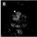
A 63 year old postmenopausal patient presented with a suspected adnexal mass. ( A ) Transvaginal ultrasound of the right adnexa with color Doppler showed a 12 cm unilocular cyst with a small amount of mural-based solid component, color score of 2 (minimal vascularity). No ascites or peritoneal nodularity is seen. This is O-RADS US 4. This patient subsequently underwent an MR imaging of the pelvis. ( B ) Sagittal T2 image and ( C ) Sagittal postcontrast subtraction image showed a corresponding unilocular cyst in the pelvis/right adnexa, with a small amount of mural-based, T2 intermediate, enhancing solid component ( arrows ). ( D ) Analysis of the dynamic contrast enhancement showed a high-risk time intensity curve, consistent with O-RADS MR imaging 5. This was pathologically proven to be endometrioid ovarian cancer.
A 39 year old premenopausal patient presented with abdominal bloating and pelvic pressure, who was found to have bilateral adnexal masses. Representative transvaginal greyscale ultrasound image of the left adnexa ( A ) shows a 9 cm multilocular, cystic mass, with a large solid component ( arrows ). Color Doppler image of the same mass ( B ) shows moderate internal flow, color score = 3. This lesion is O-RADS US 5. Subsequent pelvic MR imaging was performed. ( C ) Coronal T2-weighted image showed the corresponding left adnexal multilocular cystic mass with a solid enhancing component. The solid component shows a frond-like, branching appearance ( arrows ), the so-called “sea anemone” sign, suggesting serous papillary borderline tumor. ( D ) Axial T1 postcontrast subtraction image shows a corresponding solid component with enhancement and an intermediate-risk TIC (not shown), O-RADS MR imaging 4. There was a similar appearing right adnexal mass ( dashed arrows in C and D ). These were pathologically proven serous borderline tumors of both ovaries.
Ovarian-adnexal reporting and data system MR imaging
Related posts:
 Screening with Imaging
 Screening for Breast Cancer: Imaging Updates
 Pancreatic Cancer Screening and Early Detection
 Advancements in Screening Strategies for Early-Onset Colorectal Cancer (EOCRC)
 Prostate Cancer Screening in the MR Imaging Era
 Optimizing Screening of Hepatocellular Carcinoma with Computed Tomography and MR Imaging

Stay updated, free articles. Join our Telegram channel

Full access? Get Clinical Tree


