Ovarian Torsion
Parth C. Patel
Cassandra M. Sams
FINDINGS
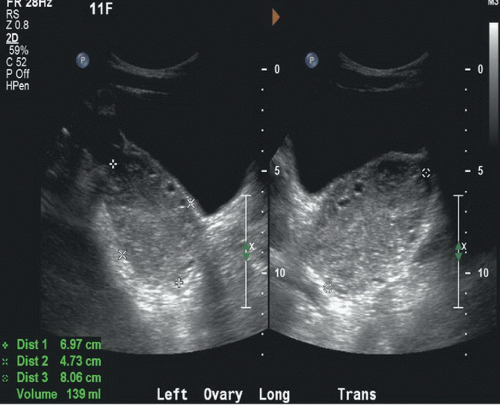
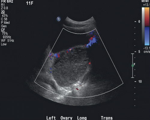
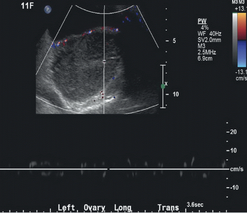
Figure 43A: Longitudinal and transverse grayscale US images show an enlarged left ovary (between cursors) with peripheral follicles. Figure 43B: Color Doppler sonogram of the left ovary shows absence of blood flow. Figure 43C: Spectral Doppler examination of the left ovary demonstrates no arterial or venous Doppler waveforms.
DIFFERENTIAL DIAGNOSIS
Ovarian mass, hemorrhagic cyst, adnexal mass, pelvic inflammatory disease.
Stay updated, free articles. Join our Telegram channel

Full access? Get Clinical Tree


