Ovarian Torsion
Parth C. Patel
Cassandra M. Sams
CLINICAL HISTORY
11-year-old female with history of acute onset of left pelvic pain.
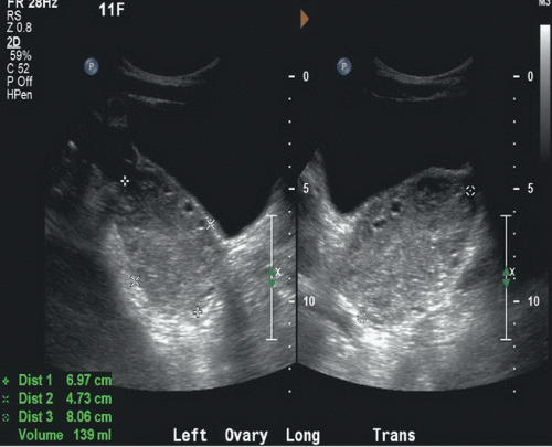 FIGURE 43A |
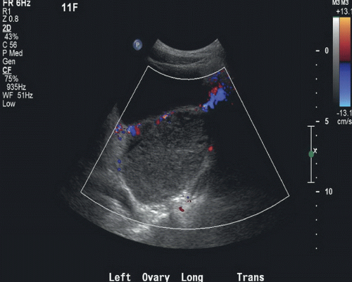 FIGURE 43B |
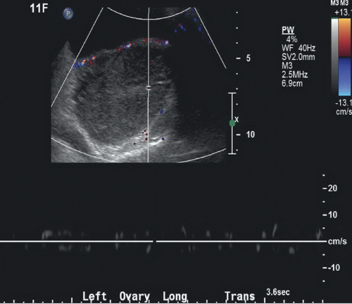 FIGURE 43C |
FINDINGS
Figure 43A: Longitudinal and transverse grayscale US images show an enlarged left ovary (between cursors) with peripheral follicles. Figure 43B: Color Doppler sonogram of the left ovary shows absence of blood flow. Figure 43C: Spectral Doppler examination of the left ovary demonstrates no arterial or venous Doppler waveforms.
DIFFERENTIAL DIAGNOSIS
Ovarian mass, hemorrhagic cyst, adnexal mass, pelvic inflammatory disease.
Related posts:


Stay updated, free articles. Join our Telegram channel

Full access? Get Clinical Tree


