Fig. 12.1
Sudan red stained aorta (left) of hyperlipidemic rabbit compared to radiograph of same after injection of 90 μCi 125I-MDA2 (right) (Reproduced with kind permission of Springer Science + Businesss Media from Tsimikas et al. [19])
This same radio-labeled antibody has also been used in subsequent experiments to demonstrate the eventual regression of atherosclerotic plaque through a modified diet [12–14].
Gamma camera scintigraphy of hyperlipidemic rabbits with 99mTc-MDA2 has also been used to successfully demonstrate areas of increased deposition of oxLDL [13] (Fig. 12.2).
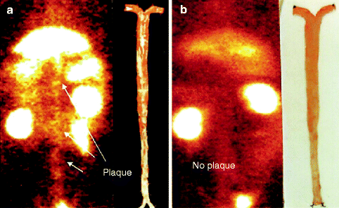

Fig. 12.2
(a, b) Scintigraphy with 99mTc-MDA2 with corresponding sudan red-stained aorta in hyperlipidemic rabbit (a) as compared to a normal rabbit (b) (Reproduced with permission of Elsevier from Tsimikas [13])
Radio-labeled (125I) native, non-modified LDL in humans successfully imaged known carotid atherosclerotic disease in humans [15].
Oxidized LDL was radio-labeled with 99mTc and used in patients who had recently suffered a transient ischemic attack. Carotid arteries with atherosclerotic plaque had significantly more uptake of the radio-labeled LDL than normal carotids [16].
Future Directions
References
1.
Choi SH, Chae A, Miller E, Messig M, Ntanios F, DeMaria AN, Nissen SE, Witztum JL, Tsimikas S. Relationship between biomarkers of oxidized low-density lipoprotein, statin therapy, quantitative coronary angiography, and atheroma: volume observations from the REVERSAL (Reversal of Atherosclerosis with Aggressive Lipid Lowering) study. J Am Coll Cardiol. 2008;52:24–32.PubMedCrossRef
2.
3.
4.
Tsimikas S, Brilakis ES, Miller ER, McConnell JP, Lennon RJ, Kornman KS, Witztum JL, Berger PB. Oxidized phospholipids, Lp(a) lipoprotein, and coronary artery disease. N Engl J Med. 2005;353:46–57.PubMedCrossRef
Stay updated, free articles. Join our Telegram channel

Full access? Get Clinical Tree


