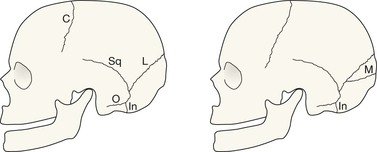
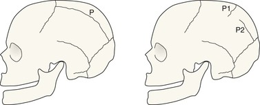
Evaluating the SXR in an infant or toddler presents unique problems. Diagnostic confusion between sutures and fractures may have serious consequences. A basic understanding of the locations and variable appearances of these sutures will help to reduce the likelihood of misdiagnosis4–8. A basic classification of skull sutures in infants and toddlers Normal accessory sutures and the radiographs on which they are seen
Paediatric skull—suspected NAI
Normal anatomy
Infants and toddlers—normal accessory sutures
Grouping
Notes
Sutures
The normal sutures
Visible on the SXR in all infants and toddlers – persisting in all adults
Sagittal, coronal, lambdoid, squamosal, and smaller sutures around the mastoid
A normal developmental suture
Visible on the SXR in all infants and many toddlers – but not in adults
Innominate
The most common accessory sutures
Visible on the SXR in some infants and toddlers – occasionally persisting to adulthood
Metopic, accessory parietal, mendosal
Suture
Most commonly seen on
Notes
Metopic suture
Frontal √ √
Towne’s √
The commonest accessory suture. It is also the one that most commonly persists in older children, and even in a few adults.
Accessory parietal suture
Towne’s √ √
Frontal √
Lateral √
May be complete or incomplete. Occurs in vertical, horizontal or oblique orientations. Most commonly vertical.
Mendosal suture
Lateral √ √
Towne’s √
Extends posteriorly from the lambdoid suture on the lateral view. Passes medially on Towne’s view.
Innominate suture
Lateral √ √
Sometimes classified as an accessory suture but best regarded as a normal developmental suture because it is always present in infants. As the child matures this suture disappears.

Paediatric skull—suspected NAI
3
