Chapter 4. Pancreas
Patient Preparation
• Fasting for 6 to 12 hours; emergency examinations may be done without fasting.
Equipment and Technical Factors
• A curved linear multihertz transducer is preferred.
• The pancreas is more echogenic than the liver in the adult patient, and isoechoic to less echogenic in pediatric age groups; images should clearly demonstrate the relational vascular landmarks.
• The pancreas generally decreases in size and increases in echogenicity with age.
• Color Doppler imaging may be used to distinguish between a vessel and a bile duct.
Imaging Protocol
• Longitudinal axis images (transverse scan plane) through the head, neck, body and tail (if possible) should be obtained; these images will also include the great vessels, portal vein, and splenic vein, and artery.
• Transverse axis images (sagittal scan plane) of the head/uncinate process, neck, body, and tail should be obtained; these images will also include the great vessels, portal vein, and splenic vein and artery.
• The relationship of the CBD and pancreas head in longitudinal and transverse axes should be demonstrated.
• Measurements of the pancreas may be included; measurements must be done when pathology is detected.
Sonographic Measurements
Pancreas
Measurements are performed in the anterior to posterior dimension perpendicular to the longitudinal axis of the pancreas:
Head: <3.0 cm (range: 2.0−3.5 cm)
Neck: 1.0−2.0 cm
Body: <2.5 cm (range: 1.2−3 cm)
Tail: <2.5 cm (range: 1.0−2.8 cm)
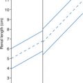
• Length: 12−15 cm (generally not measured sonographically)
• Main pancreatic duct (MPD) lumen diameter: <2 mm
| Pancreas | |||
|---|---|---|---|
| Sonographic Finding(s) | Clinical Presentation | Differential Diagnosis | Next Step |
Patient is focally tender when scanning over a pancreas with normal sonographic appearance Pancreas less echogenic than normal for age Pancreas less echogenic than normal for age and demonstrates focal or diffuse enlargement (>3.0 cm AP head or tail) With or without MPD enlargement With or without pseudocyst formation | Pain (possibly severe) Fever Fatty stool History of ERCP or pancreatic cancer Labs: elevated serum amylase (within 24 h) and lipase (within 72–94 h), direct bilirubin, ALP, WBC
Related posts:Stay updated, free articles. Join our Telegram channel
Full access? Get Clinical Tree


| ||