KEY FACTS
Terminology
- •
Solid epithelial neoplasm from ductal epithelium of exocrine pancreas
Imaging
- •
US often 1st-line imaging to evaluate obstructive jaundice and level of obstruction
- ○
Not as sensitive as CT or MR for mass or staging
- ○
- •
Hypoechoic, infiltrative mass or isoechoic mass with subtle focal contour deformity
- •
Variable size
- •
Pancreatic ductal dilation and bile duct dilation
- ○
Double duct sign: Dilation of both ducts
- ○
- •
Diffuse glandular tumor involvement can be difficult to differentiate from acute pancreatitis
- •
Atrophy or pancreatitis proximal to pancreatic ductal obstruction
- •
Typically hypovascular, but color Doppler may be helpful in assessing vascular encasement or venous obstruction
- •
Displacement/encasement of adjacent vascular structures, determines resectability (as well as metastases)
- •
Liver and regional lymph node metastases
- •
Ascites due to peritoneal metastasis
- •
Endoscopic US used to guide biopsy
Top Differential Diagnoses
- •
Chronic pancreatitis
- •
Other primary pancreatic tumors
- •
Metastases
- •
Lymphoma
Clinical Issues
- •
Mean at onset: 55 years; peak: 7th decade; M:F = 2:1
- •
Usually asymptomatic until late in course: Obstructive jaundice, weight loss
- ○
Pancreatic head tumor presents with obstructive jaundice
- ○
Body or tail tumor presents later with weight loss
- ○
- •
Most commonly presents with distant metastases (~ 65%); least likely to present with tumor confined to pancreas
- •
Poor overall prognosis; 5-year survival rate of ~ 5%
- •
Better long-term survival through complete resection
Scanning Tips
- •
Look carefully at site of transition of dilated bile or pancreatic duct to normal caliber
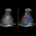
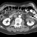
 in the pancreatic head partially obstructing the common bile duct
  and pancreatic duct
  . Superior mesenteric vessels are encased
  . Celiac nodes
  are present.
 . There are multiple liver metastases
  .
.
Stay updated, free articles. Join our Telegram channel

Full access? Get Clinical Tree


