Pancreatic Infected Necrosis and Abscess
Brooke R. Jeffrey, MD
Michael P. Federle, MD, FACR
Key Facts
Imaging
Infected pseudocyst: Often has same appearance as noninfected pseudocyst
Abscess: Well-circumscribed fluid collection, round, oval, or irregular ± gas
Necrosis: Focal, segmental, or involving entire pancreas; poorly marginated
Top Differential Diagnoses
Sterile necrosis of pancreas (necrotizing pancreatitis)
Pancreatic ductal carcinoma
Pancreatic pseudocyst
Mucinous cystic pancreatic tumor
Clinical Issues
Infected pseudocyst: Patients may be febrile but not very ill
Abscess: Patients are moderately ill
Develops late in course of acute pancreatitis (usually after 4 weeks)
Infected necrosis: Patients are very ill, “toxic”
Develops early (< 4 weeks) in course of acute necrotizing pancreatitis
CT/US-guided needle aspiration needed to establish diagnosis
Diagnostic Checklist
Distinguish among infected pseudocyst, abscess, and infected pancreatic necrosis
Infected pseudocyst: Percutaneous catheter drainage; quick recovery
Abscess: Percutaneous or surgical drainage, prolonged illness
Infected pancreatic necrosis: Surgical debridement; often repeated; high morbidity and mortality
 (Left) Axial CECT demonstrates multiple gas bubbles  within infected fluid collection in the lesser sac. (Right) Axial CECT shows extensive necrosis of the body of the pancreas with only a small residual area of enhancing parenchyma within infected fluid collection in the lesser sac. (Right) Axial CECT shows extensive necrosis of the body of the pancreas with only a small residual area of enhancing parenchyma  . Note the multiple gas bubbles . Note the multiple gas bubbles  and air-fluid level and air-fluid level  within the infected collection. There is an enlarged gastroepiploic collateral vein within the infected collection. There is an enlarged gastroepiploic collateral vein  secondary to splenic vein thrombosis. secondary to splenic vein thrombosis. |
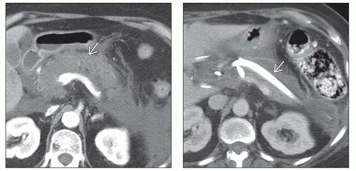 (Left) Axial CECT shows extensive necrosis with nonenhancement of the body of the pancreas  . (Right) Postoperative axial CECT in the same patient shows a large-bore drainage catheter . (Right) Postoperative axial CECT in the same patient shows a large-bore drainage catheter  in the bed of the necrotic pancreas. Despite the lack of gas bubbles on CECT, the patient underwent surgical debridement due to septicemia. Operative cultures grew multiple coliform bacteria. in the bed of the necrotic pancreas. Despite the lack of gas bubbles on CECT, the patient underwent surgical debridement due to septicemia. Operative cultures grew multiple coliform bacteria. |
TERMINOLOGY
Definitions
Infected pseudocyst: Bacterial colonization of pseudocyst
Pancreatic abscess: Circumscribed collection of pus, in or near pancreas, containing little or no pancreatic necrosis
Arises as consequence of acute pancreatitis or pancreatic trauma
Infected necrosis: Secondary infection of necrotic pancreatic tissue in setting of acute pancreatitis
IMAGING
General Features
Best diagnostic clue
Infected pseudocyst
Spherical “cyst” with enhancing wall ± gas in lumen
Abscess
Localized near-water density mass adjacent to pancreas ± gas bubbles
Infected necrosis
Poorly marginated, nonenhancing lesion in pancreas ± gas bubbles
Location
Inside or adjacent to pancreas
Size
Varies
Could be single or multiple
Morphology
Abscess
Well-circumscribed fluid collection: Round, oval, or irregular
Relatively homogeneous contents
Infected necrosis
Focal, segmental, or involving entire pancreas
Poorly marginated
Imaging Recommendations
Related posts:
Stay updated, free articles. Join our Telegram channel

Full access? Get Clinical Tree


