Pancreatic Lipomatous Pseudohypertrophy
Brooke R. Jeffrey, MD
Michael P. Federle, MD, FACR
Key Facts
Imaging
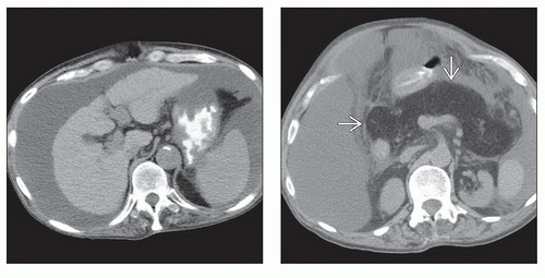
Diffuse enlargement and fatty replacement of pancreas
Without imaging or clinical signs of cystic fibrosis
Usually no evidence of ductal obstruction (pancreatic or biliary)
Top Differential Diagnoses
Cystic fibrosis
Appearance of pancreas is identical to that in lipomatous pseudohypertrophy
Clinical presentation is very different, starting in childhood
Pancreatic senescent changes
Also associated with obesity, steroid use
Pancreatic atrophy but rarely clinically evident or significant
Usually less mature-appearing fat in pancreas
Pancreatic focal fatty infiltration
Usually limited to head-uncinate or body-tail segments
Less evidence of mature fat
Shwachman-Diamond syndrome
Rare congenital multisystem disease, evident in childhood
Pathology
Associated with chronic advanced liver disease, parkinsonism, COPD
Clinical Issues
Usually incidental finding on CT or in autopsy
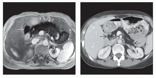
 (Left) Axial T1WI C+ FS MR in the same patient shows the lipomatous pseudohypertrophy with diffuse drop out of signal throughout the body of the pancreas
 due to fat suppression. (Right) Axial CECT in a 40-year-old man with longstanding Crohn disease shows marked fatty infiltration of the pancreas due to fat suppression. (Right) Axial CECT in a 40-year-old man with longstanding Crohn disease shows marked fatty infiltration of the pancreas  but no enlargement of the gland. The patient had no symptoms of pancreatic disease, and the fatty infiltration of the pancreas is believed to be due to chronic steroid use. but no enlargement of the gland. The patient had no symptoms of pancreatic disease, and the fatty infiltration of the pancreas is believed to be due to chronic steroid use.Stay updated, free articles. Join our Telegram channel
Full access? Get Clinical Tree
 Get Clinical Tree app for offline access

|


