Fig. 10.1


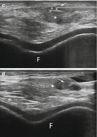
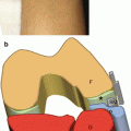
US-guided treatment of patellar tendinopathy on a short-axis scan. (a) Probe and patient position to perform short-axis US-guided treatment of patellar tendinopathy. (b) Anatomical scheme and (c) US scan of patellar tendinopathy treatment, F femur, § articular cartilage, H Hoffa’s fat pad, asterisk patellar tendon, arrow needle tip, circles peritendinous anesthesia. (d) Dry-needling procedure
Stay updated, free articles. Join our Telegram channel

Full access? Get Clinical Tree


