Edition
Publication year
Effective year
T-stage
1
1977
1978
None
2
1983
1984
T1 Small solitary tumor (<3 cm) confined to one lobe
T2 Large tumor (>3 cm) confined to one lobe, T2a: single tumor nodule, T2b: multiple tumor nodule (any size)
T3 Tumor involving both major lobes, T3a: single tumor nodule (with direct extension), T3b: multiple tumor nodules
T4 Tumor invading adjacent organs
3
1988
1989
T1 Solitary tumor 2 cm or less in greatest dimension without vascular invasion
T2 Solitary tumor 2 cm or less in greatest dimension with vascular invasion, or multiple tumors limited to one lobe, none more than 2 cm in greatest dimension without vascular invasion, or solitary tumor more than 2 cm in greatest dimension without vascular invasion
T3 Solitary tumor more than 2 cm in greatest dimension with vascular, or multiple tumors limited to one lobe, none more than 2 cm in greatest dimension, with vascular invasion, or multiple tumors limited to one lobe, any more than 2 cm in greatest dimension, with or without vascular invasion
T4 Multiple tumors in more than one lobe, or tumors involving a major branch of portal or hepatic veins
4
1992
1993
T1 Solitary tumor 2 cm or less in greatest dimension without vascular invasion
T2 Solitary tumor 2 cm or less in greatest dimension with vascular invasion, or multiple tumors limited to one lobe, none more than 2 cm in greatest dimension without vascular invasion, or solitary tumor more than 2 cm in greatest dimension without vascular invasion
T3 Solitary tumor more than 2 cm in greatest dimension with vascular, or multiple tumors limited to one lobe, none more than 2 cm in greatest dimension, with vascular invasion, or multiple tumors limited to one lobe, any more than 2 cm in greatest dimension, with or without vascular invasion
T4 Multiple tumors in more than one lobe, or tumors involving a major branch of portal or hepatic veins
5
1997
1998
T1 Solitary tumor 2 cm or less in greatest dimension without vascular invasion
T2 Solitary tumor 2 cm or less in greatest dimension with vascular invasion, or multiple tumors limited to one lobe, none more than 2 cm in greatest dimension without vascular invasion, or solitary tumor more than 2 cm in greatest dimension without vascular invasion
T3 Solitary tumor more than 2 cm in greatest dimension with vascular, or multiple tumors limited to one lobe, none more than 2 cm in greatest dimension, with vascular invasion, or multiple tumors limited to one lobe, any more than 2 cm in greatest dimension, with or without vascular invasion
T4 Multiple tumors in more than one lobe or tumors involves a major branch of portal or hepatic veins or invasion of adjacent organs other than the gallbladder, or perforation of visceral peritoneum
6
2002
2003
T1 Solitary tumor without vascular invasion
T2 Solitary tumor with vascular invasion, or multiple tumors, none >5 cm
T3 Multiple tumors, any >5 cm (T3a), or tumors involving major branch of portal or hepatic veins
T4 Tumors with direct invasion of adjacent organs other than the gallbladder, or with perforation of visceral peritoneum
7
2009
2010
T1 Single tumor without vascular invasionc
T2 Single tumor with vascular invasion, or multiple tumors, none >5 cm
T3 Multiple tumors, any >5 cm (T3a), or tumors involving major branch of portal or hepatic veins (T3b)
T4 Tumors with direct invasion of adjacent organs other than the gallbladder, or perforation of visceral peritoneum
From the sixth edition of the AJCC Cancer Staging system, the tumor size was defined as ≤ 5 cm. It was described in the sixth edition that “All solitary tumors without vascular invasion, regardless of size, are classified as T1 because of similar prognosis” [99]. However, our two large cohort studies revealed that the cumulative survival rates were significantly different among groups of patients with HCC ≤ 2 cm, 3 cm, and 5 cm [8, 18]. In addition, our published data also supported that patients with HCC above or below 3 cm had significantly different survival rates [18]. The updated BCLC also considered HCC of 3 cm to be the main factor in choice of any potentially curative option such as curative liver resection, ablation, or transplantation [100].
2.3.2 Size of HCC Versus Pathological Features
2.3.3 Size of HCC Versus Biological Behavior
Our study proposed that an HCC approaching 3 cm in diameter is reaching an important turning point for critical transformation, which changes from a relatively “benign” behavior to a more aggressive progression.[8, 18, 19, 36, 55]. This 3-cm cutoff seems to be the best point to define SHCC [8]. Other scholars have also found the relationship between tumor size and DNA ploidy [37] .
The tumor size definition in the T stage of the seventh edition of the AJCC might need to be re-evaluated by a large scale, multi-center prognostic analysis on biological stage, clinical stage, and other pathological observations. The size of HCC of 3 cm might have to be re-considered as an important factor.
2.4 Can SHCC Be Cured by RFA Basing on Its Pathological Characteristics?
Buscarini et al. in 1992 and Rossi et al. in 1993 first reported that radiofrequency ablation (RFA) is an easy-to-operate and minimally invasive technique that provides an effective local treatment. Subsequently, Shiina et al. [102] reported in cases with a small number (< 3 nodules) of small size (<3 cm in diameter) hepatocellular carcinomas (HCC), RFA was superior to the conventional HCC treatments of percutaneous ethanol injection therapy and surgical resection in terms of recurrence, complications, and survival rates. However, some tumors remain difficult to treat with RFA because these tumors cannot be visualized or are adjacent to intestinal loops or main bile ducts, which might be damaged by the treatment.
RFA can be performed percutaneously, laparoscopically, or during laparotomy and can replace surgical resection in selected patients with SHCC. However, long-term results are difficult to ascertain, because the majority of reports evaluated success in terms of tumor necrosis and few data are available on overall and disease-free survivals of patients. In this book chapter, we primarily focus on the effectiveness of RFA on SHCC based on its pathological characteristics. We also discuss protocols and new developments in ablation techniques.
2.4.1 Development of RFA in SHCC Therapy
RFA is a physical thermal ablation technique which induces thermal injury to tissues through electromagnetic energy deposition. The temperature can reach to 90–120 °C by agitation resulting in frictional heat around the electrode. This leads to immediate tissue death and thermal coagulation necrosis [103]. There are three stages of development of RFA in clinical treatment of SHCC. In the first stage (during the early 1990s), only a single and solid-center needle electrode was used, and a very small diameter of ablative region of about 1.6 cm was achieved. It was not widely adopted in clinical practice because of the limited ablative region and lack of experience in its application. It the second stage (the mid-1990s), a multiple electrode, the LeVeen electrode, and an internally cooled needle electrode were developed, which led to an increase in diameter of the ablative region to 3.5–5.0 cm. These developments made very significant improvement in the therapeutic effectiveness, and RFA gradually becomes widely used in the treatment of SHCC and other tumors. As a result, RFA gradually replaced other forms of ablative therapies and became the focus of attention. In the last stage, a new generation of electrode was invented, which integrated two-different mechanisms: a combined cluster needle electrode and a saline enhanced electrode. New electrodes used clinically for SHCC now include the expandable LeVeen (Boston Scientific Corp., Natick, MA, USA) and the monopolar Cool-tip (Covidien, Boulder, CO, USA). The LeVeen needle contains an array with diameters of 20, 30, 35, or 40 mm, and the Cool-tip needle includes a 10-mm and a 20-mm non-insulated tip, respectively. The selection of needle electrodes depends on tumor size. The selection of needle electrodes should also take into consideration the condition of the surrounding hepatic tissues to ensure a sufficient ablative margin. Masayoshi et al. [104] proposed a solution in the selection of an electrode, which can produce a wider area of ablation than what is normally required.
Over the past two decades, radiofrequency ablation (RFA) has evolved into an important therapeutic tool for treatment of SHCC. In 1996, Rossi et al. [105] first reported the long-term survival rates of RFA for SHCC. Thirty-nine patients with SHCC ≤ 3.0 cm in diameter were enrolled for RFA therapy. The overall 1-, 3-, and 5-year survival rates were 97 %, 68%, and 40 %, respectively. RFA has gradually been accepted in the treatment of SHCC, although for some clinicians, the preferred treatment for SHCC is still surgical resection and liver transplantation. RFA has significant advantages over surgery which include the following: (1) it is minimally invasive procedure and it has a high efficacy. It only takes about 10 min to ablate a tumor ≤ 3.0 cm completely. The wound is small, and the recovery is rapid; (2) it has only a relatively small impact on liver function and quality of life; (3) it is safe. A study which included 2320 patients with 3530 HCC tumors reported that the mortality after RFA was 0.3 % [106]; (4) it has a very low rate of complication; (5) it is cost-effective and has easy operability, the procedure can be done in a day clinic; and (6) the necrotic tumor tissues after treatment can become a source of autogenous vaccine, which enhances the immune response to cancer.
2.4.2 Pathological Characteristics and Other Factors Which Impact on Prognosis of RFA for SHCC
RFA offers a new option of curative treatment for SHCC. The initial results are encouraging, and it can be used in patients when surgical resection or liver transplantation is contraindicated because of poor general condition of patients. In most studies, the initial complete tumor response rates for small HCCs ≤ 3 cm following RFA have been reported to be 90–95 %. The estimated 3- and 5-year overall and disease-free survival rates were 67.0 % and 40.1 % and 68.0 and 38.0 %, respectively. The local tumor progression rates were 10–20 %. There are factors which affect good outcomes of RFA. There is no controversy that the patient’s own body mass index (BMI), which reflects technical difficulty in carrying out RFA, is significantly associated with results of RFA therapy for SHCC. Other factors include the following:
1.
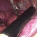
Tumor location and methods of RFA: when compared to surgical resection, percutaneous RFA is more likely to result in residual tumors, especially when the lesions are located at some specific sites of the liver, e.g., underneath the liver capsule, adjacent to the gallbladder, or under the diaphragm. Laparoscopic approach appears to be the safest and most effective method to treat small tumors on the liver surface and offers the additional advantages of laparoscopic ultrasound, which provides a good resolution to show the number and location of liver tumors. Although more invasive, open RFA can be performed and the direction of puncture of the RF needle can be better selected than the laparoscopic approach, especially for lesions located close to the gallbladder or in contact with the diaphragm.
2.
Tumor size: in a report coming from Japan, 183 patients with a solitary HCC of 3 cm or less were treated either with hepatic resection (HR) (n = 101) or RFA (n = 82) as a first-line treatment. There were no significant differences between the two groups for HCC of 2 cm or less. In patients treated with RFA, a tumor size of more than 2 cm was the only independent prognostic factor of a worse disease-free survival (risk ratio = 1.832, P = 0.039) [107].
3.
Serum albumin: Peng et al. reported that in 224 patients with a solitary HCC ≤ 5 cm and with a liver background of Child-Pugh class A treated with RFA between November 1999 and June 2007, the overall 5-, 7-, 10-year survival rates were 59.8 %, 55.2 %, 33.9 %, respectively. The median overall survival was 76.1 months. Complete ablation was achieved in 216 patients (96.4 %). Serum albumin was the only factor which significantly impacted recurrence-free and tumor-free survivals (P = 0.008, 0.002, respectively) [108].
4.
Serum alpha fetoprotein (AFP) and age: in a study from South Korea, 570 patients with 674 early-stage HCCs were treated with percutaneous RFA. The primary technique effectiveness rate was 96.7 %. The cumulative survival rates at 1, 2, 3, 4, and 5 years were 95.2 %, 82.9 %, 69.5 %, 60.8 %, and 58.0 %, respectively. Patients with Child-Pugh class A cirrhosis, younger age (≤58 years), or low AFP level (≤100 microg/L) demonstrated better survival results (P < 0.05). The Child-Pugh class, age, and AFP level before RFA were significant prognostic predictors of long-term survival [109].
5.
Endothelial cell-specific molecule 1 (ESM-1): in a study which included 150 patients with early HCC treated with RFA, ESM-1 expression by HCC stromal endothelial cells was observed in 58 patients (40 %) and it was associated with higher serum AFP levels, larger tumors, and more frequent expression of EpCAM (a surrogate marker of activation of Wnt-ß-catenin pathway). The two independent predictive factors of overall recurrence were serum AFP (HR 1.11, p = 0.045) and ESM-1 expression (HR 1.56 [1.004; 2.43], p = 0.048). Thus, ESM-1 expression was an independent predictive factor of early recurrence (HR 1.81 [1.02; 3.21], p = 0.042) [110].
6.
High serum hyaluronic acid and HBV viral load have been reported to be the main prognostic factors of local recurrence after complete radiofrequency ablation of hepatitis-B-related small HCC [111].
7.
Age: a multivariate analysis on patients with HCV-related SHCC who were treated with RFA showed age of 75 years or more [relative hazard (RH) 1.61, p = 0.019] and a serum albumin level of less than 3.5 g/dL (RH 1.61, p = 0.016), which were significant factors of a decrease in overall survival. Furthermore, a serum albumin level of less than 3.5 g/dL (RH 1.50, p = 0.003) was the only significant factor of decrease in recurrence-free survival [112].
8.
Neutrophil-to-lymphocyte ratio (NLR): an elevated preoperative NLR has been reported to be a prognostic factor for SHCC patients after RFA treatment. Multivariate analysis showed that the postoperative change in NLR, but not the preoperative NLR, was an independent prognostic factor of both overall survival (P < 0.001, HR = 2.39, 95%CI 1.53–3.72) and recurrence-free survival (P = 0.003, HR = 1.69, 95%CI 1.87–8.24). The postoperative change in NLR was an independent prognostic factor, and patients with a decrease in NLR had better survival than those with an increase in NLR [113] .
2.5 Conclusion
In conclusion, RFA is safe and effective for treating SHCC, with the advantages of having less complication, easy operation, and rapid recovery. There is no difference in disease-free survival and overall survival, and RFA and surgical resection in patients with SHCC are safe and effective. However, the pathological characteristics of SHCC with more aggressive behavior such as DNA aneuploidy, microvascular invasion, microscopic satellites, poor differentiation, capsular invasion, tumor location as well as macroscopic growth patterns, can influence the long-term treatment results for both RFA and surgical resection.
References
1.
Tang ZY. Subclinical hepatocellular carcinoma: Historical aspects and general consideration. In: Tang ZY, editor. Subclinical hepatocellular carcinoma. Berlin: Springer; 1985. p. 1–11.
2.
Zhaoyou T, Yeqin Y, Zhiying L, et al. Small hepatocellular carcinoma: clinical analysis of 30 cases. Chin Med J (Engl). 1979;92(7):455–62.
3.
Wu MC, Chen H, Zhang XH, et al. Primary hepatic carcinoma resection over 18 years. Chin Med J (Engl). 1980;93(10):723–8.
4.
Llovet JM, Bru C, Bruix J. Prognosis of hepatocellular carcinoma: the BCLC staging classification. Semin Liver Dis. 1999;19(3):329–38.PubMed
5.
Okuda K, Nakashima T, Obata H, et al. Clinicopathological studies of minute hepatocellular carcinoma. Analysis of 20 cases, including 4 with hepatic resection. Gastroenterology. 1977;73(1):109–15.PubMed
6.
Kanematsu T, Sonoda T, Takenaka K, et al. The value of ultrasound in the diagnosis and treatment of small hepatocellular carcinoma. Br J Surg. 1985;72(1):23–5.PubMed
7.
Kondo Y, Niwa Y, Akikusa B, et al. A histopathologic study of early hepatocellular carcinoma. Cancer. 1983;52(4):687–92.PubMed
8.
Cong WM, Wu MC. The biopathologic characteristics of DNA content of hepatocellular carcinomas. Cancer. 1990;66(3):498–501.PubMed
9.
Libbrecht L, Severi T, Cassiman D, et al. Glypican-3 expression distinguishes small hepatocellular carcinomas from cirrhosis, dysplastic nodules, and focal nodular hyperplasia-like nodules. Am J Surg Pathol. 2006;30(11):1405–11.PubMed
Stay updated, free articles. Join our Telegram channel

Full access? Get Clinical Tree


