, Maria Custódia Machado Ribeiro2 and Bruno Beber Machado3
(1)
Hospital da Criança de Brasília – Jose Alencar, Clínica Vila Rica, Brazília, Brazil
(2)
Hospital da Criança de Brasília – Jose Alencar, Hospital de Base do Distrito Federal, Brasília, Brazil
(3)
Clínica Radiológica Med Imagem, Unimed Sul Capixaba, Santa Casa de Misericordia de, Cachoeiro de Itapemirim, Brazil
Abstract
Many radiologists – even experienced and renowned professionals – do not feel comfortable with pediatric studies, including those related to the immature musculoskeletal system. The most important cause of this antipathy is, by far, lack of familiarity with the normal appearance of the growing skeleton and its developmental peculiarities; this unawareness is a barrier both to recognition of normal patterns and to the diagnosis of pathological findings. The purpose of this chapter is to provide the reader with a brief review of the anatomical, histological, and physiological bases of osteoarticular development, which are crucial for interpretation and understanding of pediatric imaging.
2.1 Introduction
Many radiologists – even experienced and renowned professionals – do not feel comfortable with pediatric studies, including those related to the immature musculoskeletal system. The most important cause of this antipathy is, by far, lack of familiarity with the normal appearance of the growing skeleton and its developmental peculiarities; this unawareness is a barrier both to recognition of normal patterns and to the diagnosis of pathological findings. The purpose of this chapter is to provide the reader with a brief review of the anatomical, histological, and physiological bases of osteoarticular development, which are crucial for interpretation and understanding of pediatric imaging.
2.2 The Immature Epiphysis and the Physis
A unique feature of the immature skeleton is the presence of cartilaginous structures in the extremities of the long bones, namely, the epiphyseal cartilage and the physis (also referred to as primary physis, growth cartilage, or growth plate), which involute with skeletal maturation and are no longer present in the adult organism. These cartilages act together as a functional unit responsible for the longitudinal growth of the bone. On the other hand, the articular cartilage protects and nourishes the subchondral bone of the joint surfaces, and, unlike the physis and the epiphyseal cartilage, it does not disappear, remaining throughout the life course. Radiographs are insensitive to demonstrate these cartilages (Fig. 2.1), and computed tomography and ultrasonography are also limited in their evaluation. Because of its characteristics (see Chap. 1), magnetic resonance imaging (MRI) is the preferred imaging method for assessment of most structures of the immature bone (Fig. 2.2).
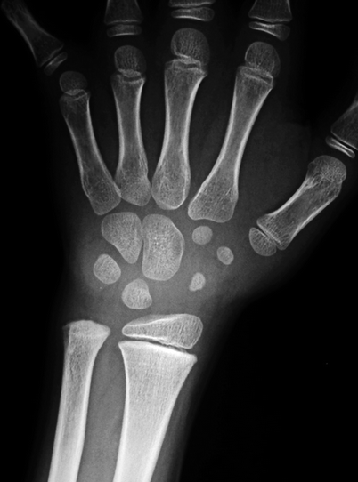
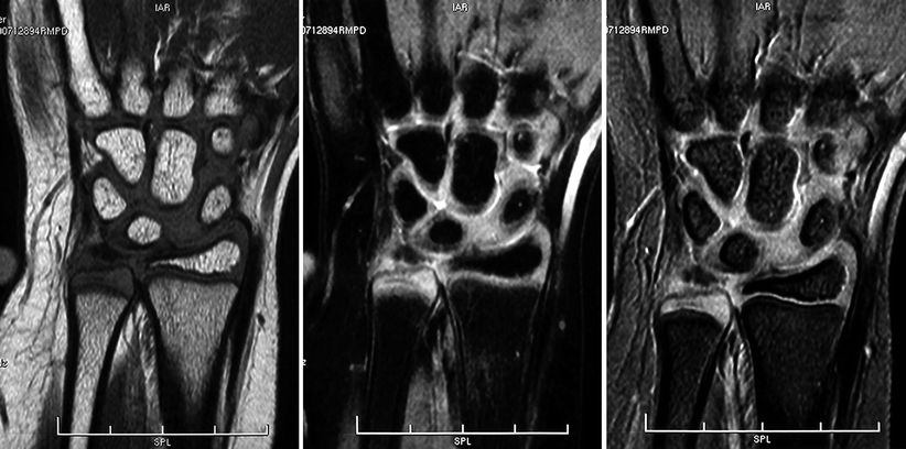
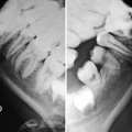
Fig. 2.1
Radiograph of the right wrist of an 8-year-old male showing that ossification has already begun in the carpal bones and in the distal radial epiphysis. However, as radiographs are fairly insensitive for cartilaginous assessment, only an indirect estimate of the thickness of the physes, of the cartilaginous anlage of the carpal bones, and of the epiphyseal cartilages can be made. The distal epiphysis of the ulna is entirely cartilaginous and completely invisible
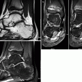
Fig. 2.2
MRI of the right wrist of a young male whose age is similar to that of the child in Fig. 2.1. Coronal T1-WI (left), fat sat PD-WI (center), and gradient-echo image (right) show that the ossified portions of the carpal bones are isointense with the medullary bone seen in the metacarpals, the distal metaphyses of the radius and the ulna, and the ossified portion of the distal epiphysis of the radius. Even though the distal epiphysis of the ulna is entirely cartilaginous, the epiphyseal cartilage is clearly seen, presenting low to intermediate signal intensity on T1-WI and intermediate to high signal intensity in the other sequences; a similar behavior is seen in the cartilaginous anlage of the carpal bones and in the cartilaginous portion of the distal epiphysis of the radius
The immature epiphysis is situated between the joint cavity and the physis. During the early stages of development, the epiphyses are entirely cartilaginous, serving as precursors for the ossified ends of the long bones. The cartilaginous epiphysis usually display low signal intensity on T1-weighted images (T1-WI) and intermediate to high signal intensity in fluid-sensitive sequences. Transformation of cartilage into bone begins in the inner portion of the epiphysis, the so-called secondary ossification center (the primary ossification centers form the diaphyses of the tubular bones during intrauterine life). Ossification may occur in a single center (like in the proximal or distal femoral epiphyses – Fig. 2.3) or, less often, via multiple secondary ossification centers that later coalesce (such as in the distal humerus – Fig. 2.4). The secondary ossification center is surrounded by a specialized structure, the secondary physis, responsible for the endochondral ossification and structurally similar to the primary physis, appearing as a thin layer of high signal intensity on fluid-sensitive sequences (Figs. 2.5 and 2.6). The ossification center, which is round when it first appears, changes to a hemispheric configuration over time: it increases in size and molds to the contour of the epiphysis in which it is settled, at the same time as there is thinning of the epiphyseal cartilage (Fig. 2.7). The epiphyseal cartilage is homogeneous throughout the first year of life, but, as skeleton maturation takes place, focal areas of high signal intensity on T2-WI appear, more evident in the posterior aspect of the femoral condyles. Such areas are more common in preadolescent males and are typically not associated with abnormalities of the subchondral bone, being deprived of significance and representing the earliest stages of endochondral ossification (Figs. 2.3, 2.4, and 2.6). They are initially focal and heterogeneous, becoming more extensive and uniform with time (Fig. 2.8). As the child begins to walk, the weight-bearing portions of the epiphyseal cartilages present a decrease in their signal intensity on T2-WI, more evident in the knees and the hips, also deprived of importance (Figs. 2.6 and 2.8). Irregularity of the secondary ossification centers at the bone/cartilage junction is a normal finding, mostly seen during growth spurts (Fig. 2.9). Accessory ossification centers may also be present, usually adjoining the posterior aspect of the secondary ossification center of the distal femur, not associated with bone marrow edema or abnormalities of the overlying cartilage. The articular cartilage is hardly discernible on T1-WI, appearing as a thin layer of high signal intensity surrounding the immature epiphysis on T2-WI (Figs. 2.5, 2.6, 2.7, and 2.10).
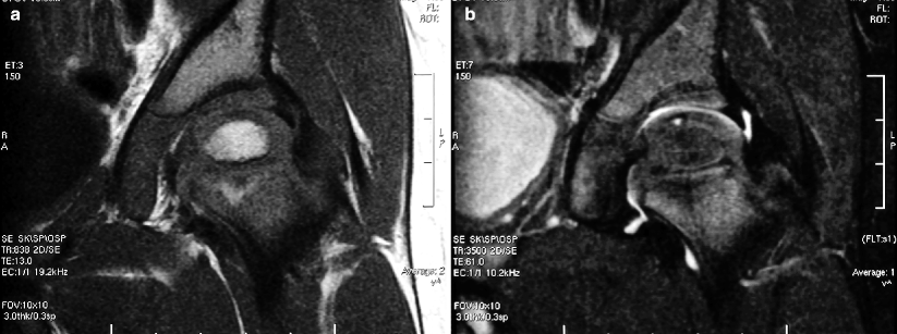
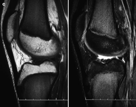
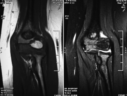
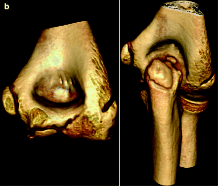
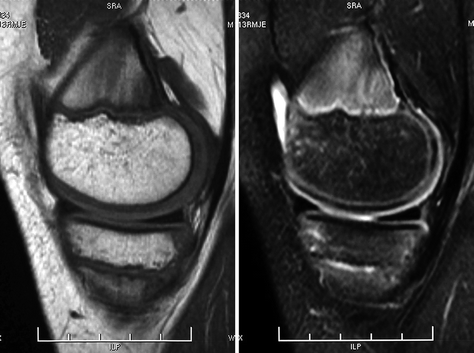
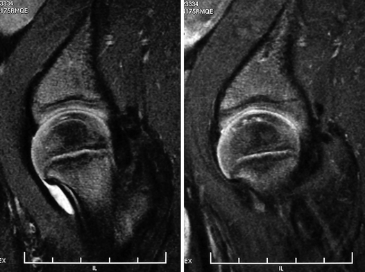
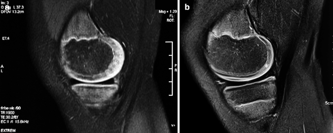
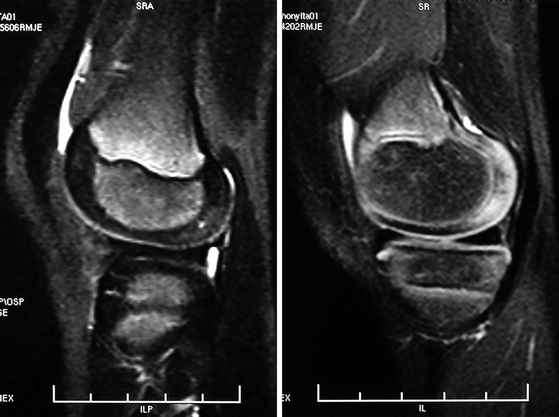
Fig. 2.3
Coronal T1-WI (a) and fat sat T2-WI (b) of the left hip of a 3-year-old male. The secondary ossification center of the femoral head is already present, whose bone marrow has undergone complete conversion to the fatty type, while the bone marrow of the femoral metaphysis and of the iliac bone presents predominance of the hematopoietic variety. The epiphyseal cartilage is predominantly isointense on T1-WI and shows intermediate signal intensity on fat sat T2-WI. There is a small focus of increased signal intensity on T2-WI in the apex of the epiphyseal cartilage, related to pre-ossification changes. The difference between the signal intensity of the yellow marrow (epiphyseal) and the red marrow (metaphyseal and diaphyseal) is even more striking in sagittal images of the knee of a school-aged child (c)
Fig. 2.4
In (a), coronal T1-WI (left) and fat sat T2-WI (right) of the left elbow of a 6-year-old female show that the ossification center of the capitulum is already filled with yellow marrow. The trochlear portion of the distal epiphysis of the humerus is completely cartilaginous, and increased signal intensity is seen in this cartilage on fat sat T2-WI, representing pre-ossification phenomena. In (b), volume-rendered CT reconstructions of the left elbow of a 13-year-old male demonstrate that all the ossification centers are ossified, with partial fusion of the ossification center of the capitulum. Despite the excellent anatomic detail of bone provided by CT, very limited information can be obtained with this imaging method about the cartilaginous structures
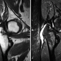
Fig. 2.5
Sagittal T1-WI (left) and fat sat T2-WI (right) of the right knee of a 13-year-old male. The secondary physis and the articular cartilage of the distal femur are clearly identified on fat sat T2-WI as linear areas of high signal intensity situated between the ossified and the cartilaginous portion of the epiphysis and along the joint surface of the epiphyseal cartilage, respectively. Bone marrow conversion is already completed in the epiphyses, while there is residual red marrow in the metaphyses, with its typical flame-shaped appearance
Fig. 2.6
Sagittal fat sat T2-WI of the left hip of a toddler. Although the epiphyseal cartilage displays predominantly intermediate signal intensity, there is low signal intensity in the weight-bearing zone, located in the uppermost portion of the epiphysis. In addition, foci of increased signal intensity are also found in the apex of the epiphyseal cartilage, related to ongoing ossification. The secondary physis appears as a thin hyperintense line surrounding the secondary ossification center, between the latter and the epiphyseal cartilage. The physis and the articular cartilage are also hyperintense if compared to the epiphyseal cartilage
Fig. 2.7
Sagittal PD-WI of the medial femoral condyles of two different children – the first is a 7-year-old subject (a) and the second one is a 13-year-old (b) – displaying the epiphyseal cartilage (*) as a structure of intermediate signal intensity between the hyperintense articular cartilage and the hypointense secondary ossification center. The ossification center is smaller, and the cartilage is proportionally thicker in younger children
Fig. 2.8
 Legg-Calvé-Perthes Disease
 Imaging Methods and the Immature Joint: An Introduction
 Sports-Related Musculoskeletal Lesions in Pediatric Patients
 Juvenile Idiopathic Arthritis
 Juvenile Spondyloarthropathies and Pediatric Collagen Vascular Disorders
 Accidental and Non-accidental Articular Injuries in the Skeletally Immature Patient


Sagittal fat sat T2-WI of the knees of two different male children, a 5-year-old (left) and a 7-year-old (right). There is rapid epiphyseal growth over time, with increase in the size of the secondary ossification center and reduction of the thickness of the epiphyseal cartilage. In the first image, marked hyperintensity is present in the primary spongiosa is of the distal femur; a zone of increased signal intensity within the posterior portion of the epiphyseal cartilage of the femoral condyle represents ongoing ossification. The second image also shows hyperintensity of the posterior portion of the epiphyseal cartilage, more diffuse if compared to that seen in the first child. The focal area of decreased signal intensity seen in the weight-bearing zone of the epiphyseal cartilage of the femur in the second image is deprived of significance, typical of ambulating children
Related posts:
Legg-Calvé-Perthes Disease
Imaging Methods and the Immature Joint: An Introduction
Sports-Related Musculoskeletal Lesions in Pediatric Patients
Juvenile Idiopathic Arthritis
Juvenile Spondyloarthropathies and Pediatric Collagen Vascular Disorders
Accidental and Non-accidental Articular Injuries in the Skeletally Immature Patient
Stay updated, free articles. Join our Telegram channel
Full access? Get Clinical Tree
