Pediatric Septic Hip
Cassandra M. Sams
CLINICAL HISTORY
5-year-old with refusal to bear weight.
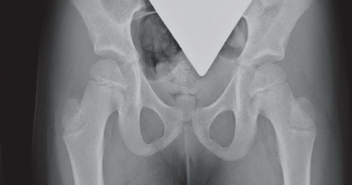 FIGURE 52A |
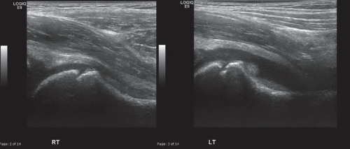 FIGURE 52B |
FINDINGS
Anteroposterior (AP) radiograph of the pelvis (Fig. 52A) is essentially normal. Sonographic images of the right and left hips obtained parallel to the long axis of the femoral neck (Fig. 52B) reveal a left hip effusion with mild thickening of the synovium; the right hip is normal.
DIFFERENTIAL DIAGNOSIS
Transient (or toxic) synovitis, inflammatory arthropathy; osteomyelitis with reactive effusion; nondisplaced fracture with associated effusion.
DIAGNOSIS
Septic arthritis.
DISCUSSION
Children presenting with hip pain are a not-infrequent sight in the emergency room. One of the most worrisome conditions to exclude is septic arthritis, a diagnosis requiring urgent treatment. In contradistinction, the diagnosis of transient synovitis is benign because it is a self-limiting phenomenon typically seen after a viral illness. Differentiating these two entities is challenging both clinically and on imaging. Laboratory data and history may be helpful in distinguishing these diagnoses with elevated inflammatory markers and WBC more suggestive of a septic process.
Related posts:


Stay updated, free articles. Join our Telegram channel

Full access? Get Clinical Tree


