Fig. 37.1
A 42 year-old female presents after motor vehicle accident. (a) Post-contrast computed tomography (CT) images of the pelvis performed during the arterial phase shows focal active hemorrhage (arrow) in the left obturator internus muscle and fracture of the adjacent superior pubic ramus (arrowhead). (b) Arterial phase image during angiography shows the site of active bleeding (arrow) from the left obturator artery. (c) On the delayed phase, more extravasated contrast (arrow) has accumulated. (d) After selection of the left obturator artery and infusion of Gelfoam slurry, a repeat angiogram shows that the bleeding has stopped
The sources of active extravasation may be either arterial or venous. With arterial injuries, the most common sources are the internal iliac artery branches. With selective catheterization (Fig. 37.1b, c) hemostasis may be achieved with embolization coils or gel foam (Fig. 37.1d).
Associated Injuries
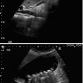
Patients with significant pelvic trauma rarely have isolated pelvic fractures. Genito-urinary injuries are particularly common with injuries to the pubic symphysis or pubic rami. In a male, a urethrogram should be performed prior to placement of a Foley catheter, particularly if blood is noted at the meatus or scrotal or perineal hematomas are identified (Fig. 37.2a, b). A cystogram should also be obtained once the Foley is placed, which can be done with plain radiographs or CT. It is important to be aware that subtle bladder ruptures may be missed if the volume of injected contrast is too small. CT cystography is far superior in the detection of subtle injuries and is preferred compared to conventional cystogram if the clinical circumstances allow. In order to prevent streak artifact the contrast dose should diluted about 1:10 in saline when performing a CT cystogram.
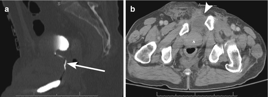
Fig. 37.2


A 74 year-old male presents after sustaining pelvic trauma. (a) Sagittal image of the pelvis during the excretory phase shows contrast filling the urinary bladder and stretching of the urethra (arrow), but no extravasation of contrast (type II injury). (b) Axial computed tomography (CT) image shows traumatic bladder extrophy (arrowhead) and diastasis of the pubic symphysis
Related posts:


Stay updated, free articles. Join our Telegram channel
Full access? Get Clinical Tree
