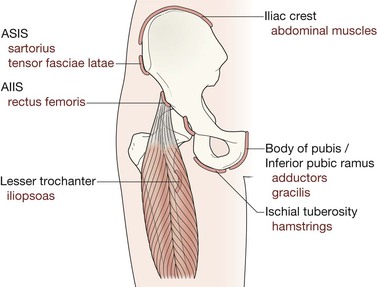
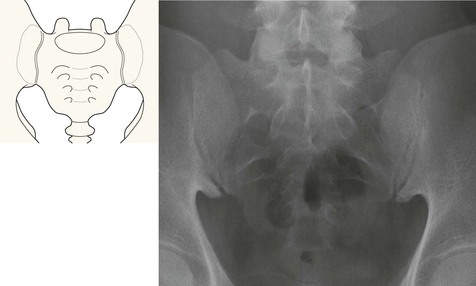
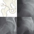
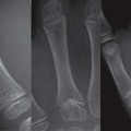
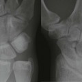
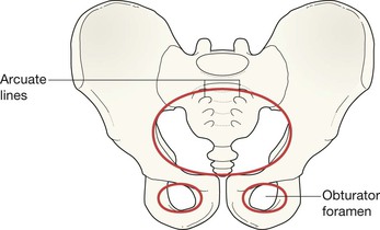
The pelvis comprises three bone rings: The robust sacro-iliac joints and the pubic symphysis are part of the main bone ring. The sacro-iliac joints are the strongest joints in the body and resist the normal vertical and anterior-posterior displacement forces; the pubic symphysis is the weakest link in the pelvic ring1–4. Arcuate lines are visible as smooth curved borders on the radiograph. They outline the roofs of the sacral formina. In children the synchondrosis (cartilaginous junction) between each ischial and pubic bone can sometimes appear confusing. In early childhood these unfused junctions may simulate fracture lines. Subsequently, between the ages of five and seven years, they may mimic healing fractures. In adolescents and young adults the pelvis shows several small secondary centres (the apophyses). These should be radiographically identical on the two sides. Apophyses are secondary centres that contribute to the eventual shape, size, and contour of the bone but not to its length. These centres are traction epiphyses as muscles originate from or insert into them. They are vulnerable to severe and acute muscle contraction, and also to repetitive forceful muscle pulls when jumping, hurdling, turning suddenly, or—occasionally—when dancing. Assess: 1. The main pelvic ring. Scrutinise both the inner and outer contours. 2. The two small rings forming the obturator foramina. 3. The sacro-iliac joints. The widths should be equal. 6. The region of the acetabulum. This is a complex area and fractures at this site are easy to overlook3,8–10. Compare the injured with the uninjured side.
Pelvis
Normal anatomy
Normal AP view
Developing skeleton: Synchondroses

Developing skeleton: Pelvic bone apophyses5–7
Analysis: the checklist
The AP radiograph
In adolescents and young adults presenting with hip pain but no history of a violent blow also assess:

Pelvis
13
