, Stefano Fanti1 and Lucia Zanoni1
(1)
Department of Nuclear Medicine, Universitary Hospital Sant’Orsola-Malpighi, Bologna, Italy
Endometrial Carcinoma
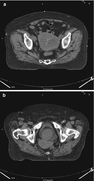
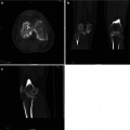
Fig. 5.1
(a, b) Huge mass in the endometrium involving the uterine body
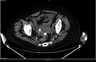
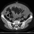
Fig. 5.2
Endometrial and ovary cancer in a breast cancer patient
CT findings in endometrial cancer include:
Relatively hypo-attenuated mass in the region of the endometrial cavity which may show uniform attenuation or may be heterogeneous, with or without a contrast-enhanced component
Polypoid mass surrounded by endometrial fluid
Heterogeneous soft-tissue mass/masses and fluid expanding the endometrial cavity
Tumor involving multiple regions of the endometrium or the entire endometrial surface
Fluid-filled uterine cavity marginated by mural tumor implants
Fluid-filled uterine cavity secondary to obstruction of the endocervical canal by tumor that is not depicted or delineated clearly.
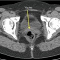
Groin Lymphadenopathy
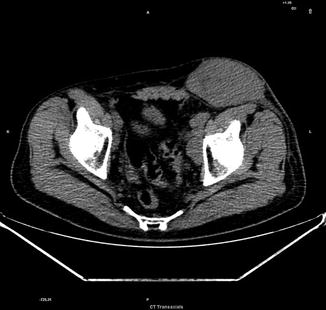
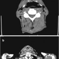
Fig. 5.3
Left groin mass suggestive for lymphoma
Related posts:

Stay updated, free articles. Join our Telegram channel

Full access? Get Clinical Tree


