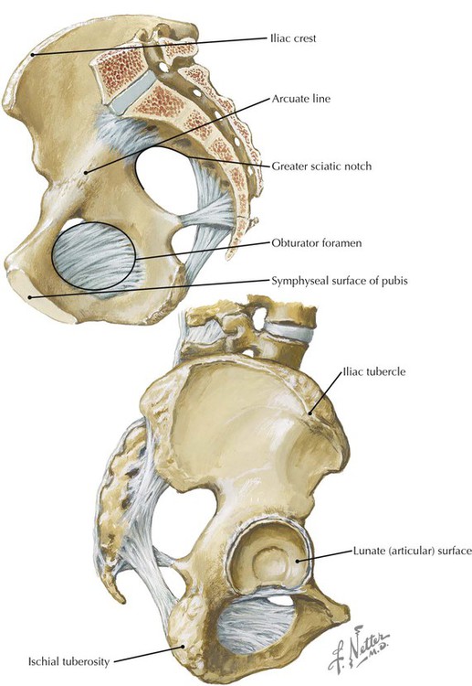
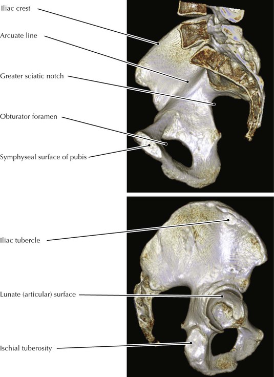
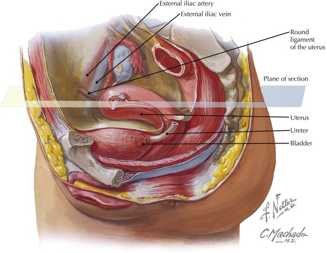
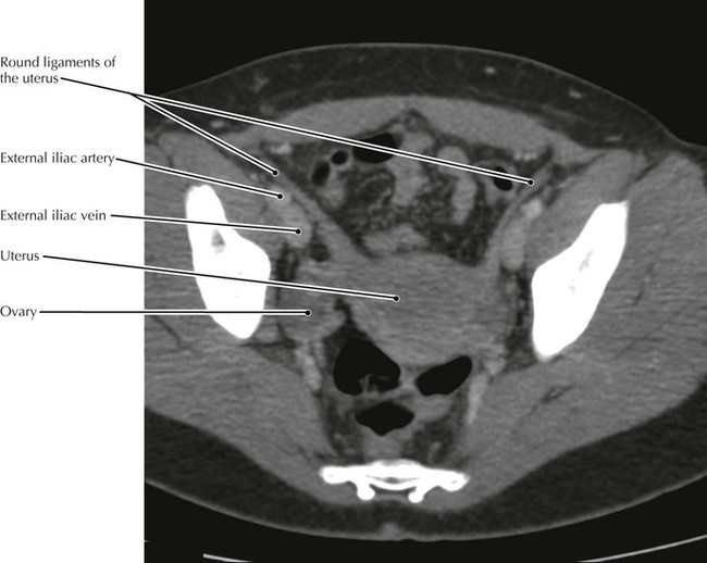
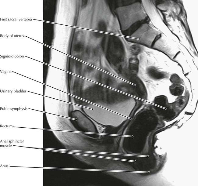
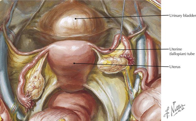
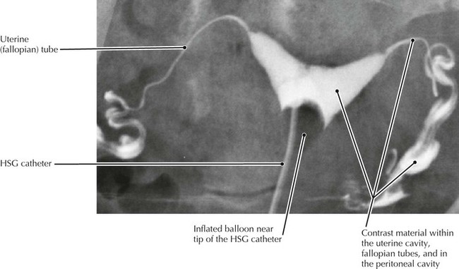
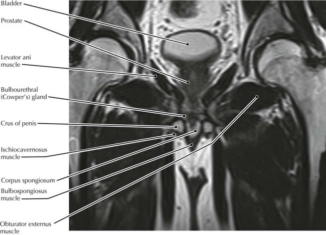
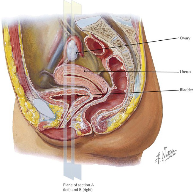
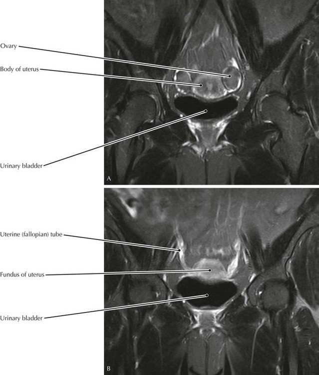
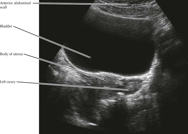
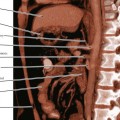
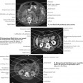
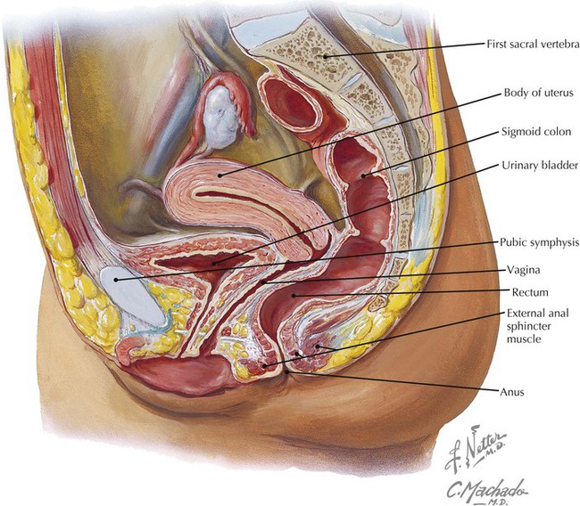
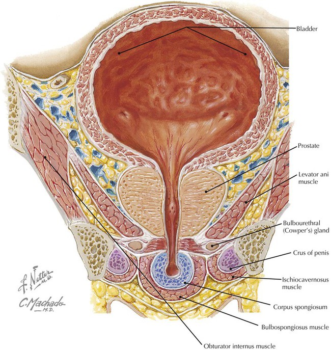
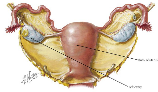
• The external surface of the iliac blade is the attachment site for the gluteus medius and minimus muscles. These two muscles are primarily responsible for maintaining pelvic stability when one foot is lifted off the ground (e.g., during the swing phase of walking). They both insert into the greater trochanter of the femur. • The lunate surface is the articular part of the acetabulum. • The symphyseal surface of the pubis undergoes predictable changes with aging so that the surface can be used to estimate the age of skeletal material collected forensically or archeologically. • The round ligament passes through the inguinal canal to reach the labia majus. Lymph vessels travel with the ligament so that some lymph from the uterus drains to the inguinal nodes. • The position of the ovary can vary within a patient with the amount of bladder and bowel distention and the patient’s position. • The anal sphincter has both internal (innervated by pelvic splanchnic [parasympathetic] nerves) and external (innervated by the inferior rectal [somatic] nerves) divisions. • The uterus is not well seen in this patient because much of it extends to one side, out of the plane of this midline sagittal image. Such “tilting” and other variations of the uterus are common and must be kept in mind when viewing cross-sectional images so that erroneous conclusions pertaining to uterine conditions are not made. • The levator ani muscle comprises most of the pelvic diaphragm and is critical to the maintenance of urinary and fecal continence. • The bulbospongiosus muscle is a sphincter of the urethra and may play a role in maintaining an erection by forcing blood into the distal penis. • The ischiocavernous muscles also function in that manner to maintain an erection.
Pelvis and Perineum
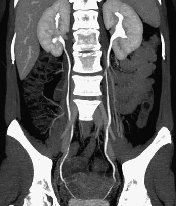
Pelvis
Female Pelvis, Round Ligament, and Ovary
Female Pelvic Viscera, Sagittal View
Bulb of Penis, Coronal Section
Uterus and Adnexa
Pelvis and Perineum

