Pericarditis
Sam A. Glaubiger
CLINICAL HISTORY
10-year-old female with chest pain and fever.
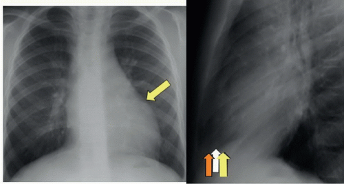 FIGURE 50A |
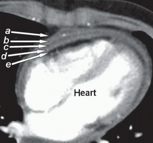 FIGURE 50B |
FINDINGS
Figure 50A: Posteroanterior (PA) chest film (left) shows some fullness of the left cardiac border (yellow arrow). Lateral chest film (right) shows subtle differences in density along the anterior heart border. Fluid in the pericardial space (white arrow) is more dense than pericardial fat (orange arrow) and more dense than epicardial fat (yellow arrow). These tissues together account for the layered appearance or “sandwich” sign of pericarditis and/or pericardial effusion. Figure 50B: Contrast-enhanced axial CT image of the heart. Pericardial fat (a) and epicardial fat (e) are on either side of thickened parietal (b) and visceral (d) pericardium with a small pericardial effusion (c). The pericardial layers are all the same density on plain film, composing the middle of the “sandwich.”
Related posts:


Stay updated, free articles. Join our Telegram channel

Full access? Get Clinical Tree


