Perilunate Dislocation
Cody J. Schwartz
Daniel B. Nissman
CLINICAL HISTORY
28-year-old female with a history of motor vehicle collision (MVC) who presents with wrist pain.
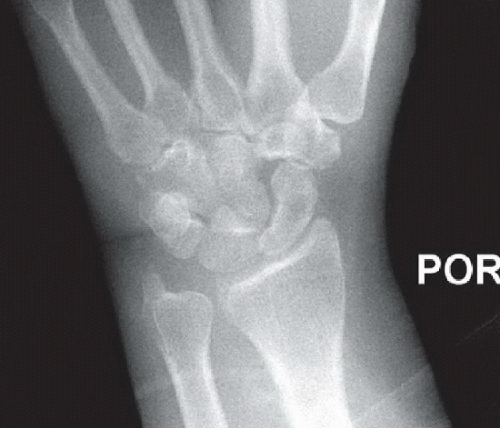 Figure 32A |
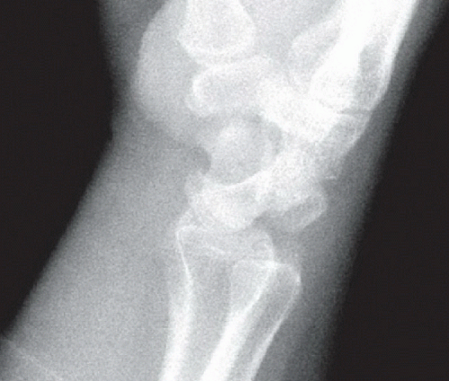 Figure 32B |
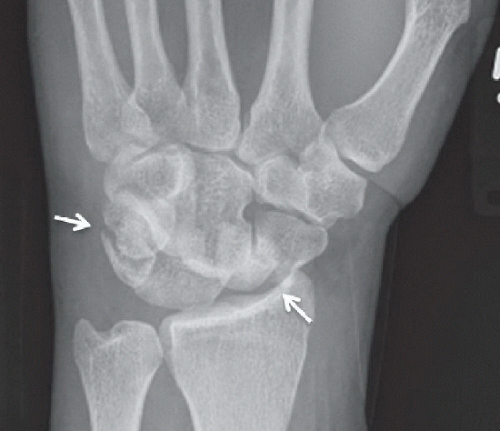 Figure 32C |
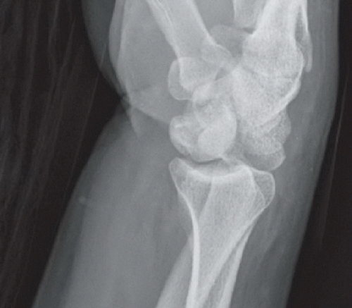 Figure 32D |
FINDINGS
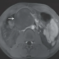
Portable posteroanterior (PA) radiograph of the left wrist (Fig. 32A) shows a triangular lunate with abnormal overlap of the lunate and capitate (“piece of pie” sign). The middle carpal arc (distal concave curve of the scaphoid, lunate, and triquetrum) is disrupted. Subtle contour abnormality of the proximal carpal arc (proximal convex curve of the scaphoid, lunate, and triquetrum) at the scapholunate joint is also noted. The portable lateral radiograph of the wrist (Fig. 32B) shows, with the exception of the lunate, dislocation of the entire carpus dorsal to the radius; only the lunate maintains its normal relationship with the radius. PA (Fig. 32C) and lateral (Fig. 32D) radiographs of the wrist from a different patient show similar findings with additional scaphoid waist and triquetral fractures (arrows).
DIFFERENTIAL DIAGNOSIS
Lunate dislocation, perilunate dislocation, lunate fracture-dislocation, perilunate fracture-dislocation.
DIAGNOSIS
Dorsal perilunate dislocation (Figs. 32A and 32B), dorsal transscaphoid transtriquetral perilunate fracture-dislocation (Figs. 32C and 32D).
Related posts:


Stay updated, free articles. Join our Telegram channel

Full access? Get Clinical Tree


