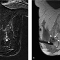
9 Perioperative Specimen Imaging When an excisional biopsy is performed for a nonpalpable breast lesion, imaging must verify that the suspicious lesion has been removed and biopsy is representative. This is not usually necessary for palpable findings because it can be assumed that the surgeon is able to reliably excise these. Which imaging technique to perform for this perioperative check is dependent upon whether the biopsied lesion is most reliably identified on mammography (specimen radiography), or ultrasound (US) (specimen US). Specimen magnetic resonance imaging (MRI) has not been established as a reliable method to verify correct excision. Good Practice Recommendation* Preoperative localization and postoperative specimen imaging should always be performed when a nonpalpable breast lesion is excised (Grade of Recommendation A, Level of Evidence 3b). Mammography of surgically excised tissue—a specimen radio-graph—is essential after open surgery of mammographic abnormalities associated with microcalcifications. Pertinent information derived from the specimen radiograph is whether any microcalcifications are detected at all, whether the biopsy is representative and/or the microcalcifications completely excised, and whether the microcalcifications are centrally or marginally located in the specimen. A specimen radiograph is also obligatory after surgical removal of nonpalpable breast lesions. The target abnormality can often be radiographically detected within the specimen, especially when the surrounding tissue is lipomatous. In such cases, the specimen radiograph is a reliable documentation of the representative lesion excision. Examination in two planes. Specimen radiography should be performed in two orthogonal planes (craniocaudal [CC] and mediolateral [ML]) (Fig. 9.1). This can be done by placing the specimen in a commercially available container (e. g., a Bollmann specimen chamber) or in any other suitable, x-ray transparent container (Fig. 9.2). Some containers, however, require that the specimen be repositioned for radiography, occasionally making topographic orientation more difficult. Others take anatomic relationships with regard to the axilla and other defined landmarks into consideration. Fixation. The assignment of the specimen configuration to the anatomic topography is facilitated when the surgical specimen is fixed or mounted on a carrier medium, and can be x-rayed on this medium without further manipulation. To facilitate this, the materials used must allow the specimen to be firmly fastened, as well as be easily x-rayed without producing disturbing artifacts. One material possessing these qualities is Styrofoam. Other materials, such as cork, often possess interfering, radiopaque inclusions (Fig. 9.3). As an aid to the pathologist, it is also possible to place markers on relevant areas of the tissue specimen as seen on the specimen radiograph and then take another radiograph for documentation (Fig. 9.4).
Specimen Radiography
Stay updated, free articles. Join our Telegram channel

Full access? Get Clinical Tree


