Peritonitis
R. Brooke Jeffrey, MD
Key Facts
Terminology
Infectious or inflammatory process involving peritoneum or peritoneal cavity
Imaging
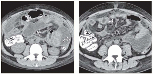
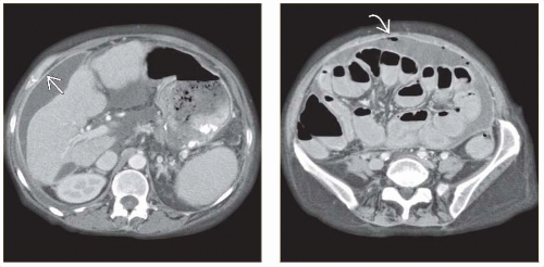
Best imaging clue: Ascites, symmetric enhancement of peritoneum with abdominal fat stranding on CECT
Peritoneal fluid, septations, thickened echogenic mesentery on US
Best imaging tool: CECT or US
Top Differential Diagnoses
Peritoneal carcinomatosis
Benign ascites
Pseudomyxoma peritonei
Hemoperitoneum
Pathology
Pus in peritoneal cavity, thickened peritoneum or mesentery
Clinical Issues
Most common signs/symptoms: Fever, abdominal pain, distension
Diagnostic Checklist
Consider peritoneal carcinomatosis
Imaging pearls
Symmetric enhancement of thickened peritoneum
Enlarged fallopian tube with fluid-fluid level and complex adnexal mass in PID
Inflammatory changes with adjacent fat of mesentery and omentum
TERMINOLOGY
Definitions
Infectious or inflammatory process involving peritoneum or peritoneal cavity
IMAGING
General Features
Best diagnostic clue
Ascites, symmetric enhancement of peritoneum with abdominal fat stranding
Location
Peritoneal surface, mesentery, omentum
Size
Variable, may be focal or diffuse
Morphology
Symmetric thickening of peritoneum
Radiographic Findings
Radiography
Evidence of ascites: > 500 mL fluid required for plain film diagnosis
Flank bulging
Indistinct psoas margin
Small bowel (SB) loops floating centrally
Lateral edge of liver displaced medially (Hellmer sign); visible in 80% of patients with significant ascites
Pelvic “dog’s ear” present in 90% of patients with significant ascites
Medial displacement of cecum and ascending colon present in 90% of patients with significant ascites
± free air
Hydropneumoperitoneum
Air in lesser sac with perforated gastric ulcer
Fluoroscopic Findings
Upper GI
Perforated ulcer with contrast leak
Contrast enema
Diverticular perforation in diverticulitis
CT Findings
CECT
Ascites, enhancing peritoneum with smooth thickening, infiltration, and soft tissue fat stranding within mesentery
± gas bubbles, low-attenuation nodes in TB peritonitis
MR Findings
T1WI
Low-signal peritoneal fluid
T2WI
High-signal peritoneal fluid
T1WI C+
Stay updated, free articles. Join our Telegram channel

Full access? Get Clinical Tree


