Fig. 1
Newly diagnosed malignant fibrous histiocytoma. From left to right, a computed tomography (CT), b positron emission tomo — graphy (PET), c fused PET-CT and d maximum intensity projection PET show the heterogeneous appearance of the mass with large areas of necrosis. These data can guide the biopsy site to areas showing increased 18F-fluorodeoxyglucose uptake (for color reproduction see p 310)
Poor risk factors in patients with STS include highgrade tumor, large tumor size (over 5 cm in largest diameter), tumor necrosis, deep tumor location, proximal location in tumors in the lower extremity, metastatic spread, failure to achieve tumor-free margin after surgical resection, and local recurrence [1].
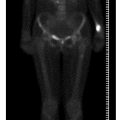
PET-CT, CT, bone scintigraphy and radiography are often used as complementary modalities to assess the presence of metastases. Metastatic spread of sarcomas is mainly hematogenous, although lymphatic spread may occur [2]. Lung followed by bone are the most common sites of STS metastasis. 18F-FDG PET-CT whole body imaging is a useful modality for staging and detecting distant metastasis, with a high sensitivity and specificity and excellent negative predictive value [5]. When interpreting 18F-FDG PET-CT images, both functional data of PET and morphological data of CT should be considered. The latter is valuable mainly for detection of lunginvolvement, since PET may be falsely negative in the presence of small lung metastases identified by CT [5]. As for other tumors, the standard 99mTc-methylene diphosphonate (MDP) bone scan (BS) is widely used for assessment of skeletal involvement in STS, detecting bone metastases of osteoblastic type. 18F-FDG PET-CT is more accurate for detection of lytic type metastasis and early marrow involvement, for which BS is insensitive [7]. In a recent study, detection of unexpected metastasis by 18F-FDG PET-CT was reported to result in a change in treatment for 18 of 59 STS patients, mainly cancellation of surgery and referral for chemotherapy instead [5].
Patients with low-grade small STS often undergo surgery and occasionally adjuvant radiation. Surgical resection ranges from simple excision with tumor-free wide margins to complex limb salvage procedures. Patients with large intermediate-grade or high-grade large tumors are usually referred for neoadjuvant chemotherapy, with or without preoperative radiation, followed by resection and adjuvant therapy. Based on percentage of tumor necrosis in the resected tumor, a regimen of adjuvant therapy is selected so that if an adequate response is not achieved, an alternative chemotherapy regimen replaces the ineffective treatment. Chemotherapy resistance is a crucial prognostic factor. The complexity and genetic and chromosomal instability found in STS are probably the grounds for the high incidence of multidrug resistance and consequent treatment failure [16, 17].
Monitoring response to therapy is a principle role of 18F-FDG PET-CT in patients with STS particularly since the RECIST (response evaluation criteria in solid tumors) criteria for treatment response do not apply well in this cohort, as successful treatment often results in larger areas of inflammation and necrosis and no signif icant decrease in the tumor size [18–20]. Decrease in tumor metabolism reflected by decrease in 18F-FDG uptake has been shown to precede tumor size reduction in association with therapeutic success [1]. The novel biological anticancer drugs used in some types of sarcoma are often associated with lag in tumor shrinkage. Knowledge of the timing of performance of the PET-CT study in relation to the course and type of treatment is of great importance when interpreting the findings. When performed early after initiation of therapy, the primary clinical demand of imaging is to assess the cytostatic effect of chemotherapy in order to exclude nonresponders or modify the therapeutic protocol accordingly. Decrease or no uptake of 18F-FDG during or immediately after initiation of therapy might reflect metabolic shutdown but not necessarily death of tumor cells. As shown in a recent study on 39 STS patients, 18F-FDG PET-CT can predict survival soon after the initial cycle of neoadjuvant chemotherapy, and thus may be used as an intermediate endpoint biomarker [21].
When performed after completion of therapy, a negative 18F-FDG PET suggests no active disease even in the presence of a residual mass, as the latter can be composed of hemorrhage, edema, necrosis or scar with no active tumor tissue [8, 22]. It should be borne in mind that early after radiotherapy, inflammatory cells of granulation tissue may show increased 18F-FDG uptake with potential false-positive interpretation. Evilevitch et al. [18] investigated 42 patients with biopsy-proven high-grade STS who underwent 18F-FDG PET-CT scans before and after neoadjuvant chemotherapy. Metabolic parameters were correlated with RECIST criteria as well as histopathologic response. A decrease in 18F-FDG uptake of greater than 60% showed all histopathologic responders with sensitivity of 100% and specificity of 71%, whereas application of RECIST criteria resulted in a sensitivity of 25% and a specificity of 100%.
Approximately 10–15% of STS patients develop a local recurrence that might be difficult to identify on morphologic imaging modalities because of anatomy distorted by previous surgery and radiotherapy [2]. 18F-FDG PET-CT was found to be valuable for detection of local recurrence despite treatment-induced altered anatomy [23].
18F-FDG PET/CT imaging for GIST treatment response evaluation has been incorporated into the guidelines for GIST management [24]. In responders, a rapid and almost complete shutdown of glucose metabolism, reflected by decrease or disappearance of 18F-FDG uptake, is observed almost immediately after the start of imatinib mesylate treatment preceding a potential change in tumor size on CT in several weeks, a change that is not always present, even when treatment is successful [25]. Early metabolic response of GIST is associated with a significant longer progression-free survival [9].
In summary, 18F-FDG PET/CT is a beneficial staging and follow-up imaging modality in this cohort. However, there is no consensus as to its introduction in the imaging algorithm nor standardization of the criteria for monitoring response to therapy. Such consensus requires a large amount of accumulated clinical data for each of the STS subtypes and long enough clinical follow-up. As PET-CT has been used for over a decade now, I believe that standardization and guidelines are soon to come.


