Abstract
Rationale and objectives
Recent literature suggests that performing systematic head computed tomography (CT) scans for mild traumatic brain injury (mTBI) in patients undergoing antithrombotic therapy offers limited benefits. This study aims to evaluate a set of criteria that could potentially eliminate the need for systematic head CT scans, performed solely because of the antithrombotic treatment status, in elderly patients (aged 75 or older) presenting with mTBI.
Materials and methods
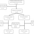
All patients aged 75 or older who underwent a head CT scan at our academic center for mTBI while on antithrombotic therapy between January and December 2022 were retrospectively included in this study. Patients were categorized into two groups. The first group, referred to as the “At-risk group”, included patients with any of the following: GCS score < 15 or cognitive impairment; initial loss of consciousness; hemodynamic instability; signs of fractures; extensive subcutaneous hematoma; severe or treatment-resistant headache; vomiting; seizure; any neurological deficit; intoxication; amnesia; or a history of neurosurgery. The second group, referred to as the “Not-at-risk group”, comprised patients without any of these criteria.
Results
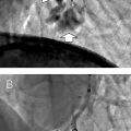
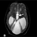
A total of 1415 patients were included. Post-traumatic intracranial hemorrhage ( P < 0.001), brain herniation ( P = 0.003), and fractures ( P < 0.001) occurred statistically more frequently in the At-risk group. Six post-traumatic hemorrhagic brain injuries were found in the Not-at-risk group, that did not present any of the studied criteria, and all these injuries were minor (localized SAH; millimetric SDH). Furthermore, none of these required immediate or delayed surgical intervention, and no neurological deterioration or deaths occurred in these patients.
Conclusion
In conclusion, conducting systematic head CT scans based solely on antithrombotic therapy in elderly patients aged 75 or older with mTBI might be irrelevant.
mTBI
mild traumatic brain injury
GCS
Glasgow coma scale
CT
computed tomography
mRS
modified Rankin scale
PACS
picture archiving and communication system
SD
standard deviation
IQR
interquartile range
SPSS
Statistical Package for the Social Sciences
1
Introduction
Mild traumatic brain injury (mTBI) refers to an acute brain injury resulting from the transfer of mechanical energy to the head by external physical forces, with a Glasgow Coma Scale (GCS) score following mTBI ranging from 13 to 15. It encompasses impacts to the head as well as acceleration and deceleration movements without direct trauma to the skull. It is estimated that globally, 90 % of all head injuries are mTBIs, making them a common reason for visits to emergency departments [ ].
The management of traumatic brain injury represents a significant socio-economic cost, amounting to nearly 400 billion dollars annually worldwide [ ]. The transfer of a patient with mTBI to a hospital facility equipped with an emergency department, along with the use of computed tomography (CT) for diagnostic purposes, contributes to higher public health costs. A reduction in the number of CT scans performed in the aftermath of a traumatic brain injury would constitute a major medical-economic saving [ , ]. mTBIs disproportionately impacts the elderly, especially those over the age of 75, who represent a unique patient demographic. Within this population, injuries frequently result from low-kinetic events, most commonly falls from standing height [ ]. Additionally, the options for surgical intervention are constrained for these patients [ ]. Recognizing that moving an elderly person from their usual living environment can exacerbate neurological issues, the practice of transferring mTBI patients for routine emergency imaging is subject to debate [ ]. This concern is particularly relevant for individuals living in institutional settings, such as retirement or nursing homes, where while supervision is provided, there may be an absence of on-site facilities capable of conducting brain CT scans.
Recent literature suggests that performing systematic head CT scans for mTBI in patients undergoing antithrombotic therapy offers limited benefits, particularly in adults younger than 65 years of age [ , ]. However, the necessity of such diagnostic procedures in elderly patients remains uncertain. This study aims to evaluate a set of criteria that could potentially eliminate the need for systematic head CT scans, performed solely because of the antithrombotic treatment status, in elderly patients (aged 75 or older) presenting with mTBI.
2
Materials and methods
2.1
Study design and patient characteristics
All patients who underwent a head CT scan at our academic center for mild traumatic brain injury while on antithrombotic therapy are included in a database. Patients who underwent head CT scan between January 2022 and December 2022 were retrospectively included in this study using this database.
The inclusion criteria were as follows: patients aged ≥ 75 years, treated with antithrombotics (either anticoagulants or antiplatelets), who presented at the emergency department within 24 h after trauma, with a GCS score of 13–15. The exclusion criteria were as follows: age < 75 years, GCS < 13, transfer from other hospitals, high-impact trauma (high kinetics).
Clinical characteristics of the patients were documented from both paper medical records and digital files. Information gathered included age, sex, location of the trauma (such as own residence, retirement home, nursing home, or hospital), modified Rankin scale (mRS) score, type of antithrombotic medication.
Patients were categorized into two groups, with criteria inspired by current guidelines from France, Canada, the United Kingdom, and the United States [ , ]. The first group, referred to as the “At-risk group”, included patients with any of the following, either alone or in combination: a GCS score < 15 or cognitive impairment; initial loss of consciousness; hemodynamic instability; signs of skull vault, skull base, or facial fractures (including otorrhagia, rinorrhagia, spectacle hematoma, etc.); extensive subcutaneous hematoma; severe or treatment-resistant headache; vomiting; seizure; any neurological deficit; drug or alcohol intoxication; amnesia related to the event; or a history of neurosurgery. The second group, referred to as the “Not-at-risk group”, comprised patients without any of these criteria.
Outside of the initial visit to the emergency department, which could occasionally be followed by hospitalization within the same institution, no systematic follow-up protocol was implemented. Consequently, data on patient outcomes beyond this period were not routinely collected.
2.2
CT scan protocol and image analysis
CT scans were conducted without the injection of contrast medium, utilizing volume spiral acquisition from the cranio-cervical junction to the vertex, and a section thickness of 1.25 mm. The studies from all patients were placed in an anonymized folder on the picture archiving and communication system (PACS). The CT scans were independently reviewed by a neuroradiology resident and a senior neuroradiologist. In cases of disagreement, a consensus was reached between the two radiologists.
The elements gathered included the presence or absence of intracranial hemorrhage; the type of hemorrhage, categorized into petechial hemorrhages, parenchymal hematoma, subarachnoid hemorrhage, extradural hematoma, subdural hematoma, and intraventricular hemorrhage; presence or absence of brain herniation; type of herniation; and the presence or absence of skull vault, skull base, or facial fractures, along with the type of fracture.
Due to the retrospective nature of this study and missing data, it was not possible to determine the precise average time interval between trauma and the completion of the CT scan.
2.3
Statistical analysis
Continuous data are presented as mean (standard deviation [SD]) or median (interquartile range [IQR]). Categorical variables are presented as count (percentage). Statistical comparisons were performed by a Student t -test for normally distributed data, the Mann-Whitney U test for data with a skewed distribution, and the ꭓ² and Fisher’s exact tests for categorical data. A P value < 0.05 was statistically significant. The data were analyzed using the Statistical Package for the Social Sciences (SPSS; Version 28.0.1.1, IBM, Armonk).
3
Results
3.1
Patient characteristics
Table 1 presents the descriptive data for the study population, categorized according to the two groups previously defined.
| Total ( n = 1415) | At-risk group ( n = 772, 54.6 %) | Not-at-risk group ( n = 643, 45.4 %) | P | |
|---|---|---|---|---|
| Age (years), median (IQR) | 87 (82–91) | 87 (82–91) | 87 (82–91) | 0.613 |
| Women, count (percentage) | 833 (58.9 %) | 459 | 374 | 0.623 |
| Location of the trauma, count (percentage) | ||||
| – Own residence | 999 (70.6 %) | 529 (68.5 %) | 470 (73.1 %) | 0.06 |
| – Retirement home | 84 (5.9 %) | 43 (5.6 %) | 41 (6.4 %) | 0.523 |
| – Nursing home | 211 (14.9 %) | 143 (18.5 %) | 68 (10.6 %) | < 0.001 |
| – Hospital | 121 (8.6 %) | 57 (7.4 %) | 64 (10.0 %) | 0.085 |
| Initial mRS, median (IQR) | 3 (3–3) | 3 (3–3) | 3 (3–3) | < 0.001 |
| Antiplatelet therapy, count (percentage) | 729 (51.5 %) | 398 (51.6 %) | 331 (51.5 %) | 0.977 |
| Anticoagulant therapy, count (percentage) | 722 (51.0 %) | 390 (50.5 %) | 332 (51.6 %) | 0.676 |
| Other hemostasis issue, count (percentage) | 7 (0.5 %) | 1 (0.1 %) | 6 (0.9 %) | 0.051 |
| Criteria related to the trauma (and assessed in the present study) , count (percentage) | ||||
| – GCS < 15 or cognitive impairment | 474 (33.5 %) | 474 (61.4 %) | 0 (0.0 %) | NA |
| – Initial loss of consciousness | 89 (6.3 %) | 89 (11.5 %) | 0 (0.0 %) | NA |
| – Hemodynamic instability | 6 (0.4 %) | 6 (0.8 %) | 0 (0.0 %) | NA |
| – Sign of fracture | 100 (7.1 %) | 100 (13.0 %) | 0 (0.0 %) | NA |
| – Extensive subcutaneous hematoma | 175 (12.4 %) | 175 (22.7 %) | 0 (0.0 %) | NA |
| – Severe or treatment-resistant headache | 29 (2.0 %) | 29 (3.8 %) | 0 (0.0 %) | NA |
| – Vomiting | 26 (1.8 %) | 26 (3.4 %) | 0 (0.0 %) | NA |
| – Seizure | 8 (0.6 %) | 8 (1.0 %) | 0 (0.0 %) | NA |
| – Neurological deficit | 15 (1.1 %) | 15 (1.9 %) | 0 (0.0 %) | NA |
| – Drug or alcohol intoxication | 16 (1.1 %) | 16 (2.1 %) | 0 (0.0 %) | NA |
| – Amnesia related to the event | 92 (6.5 %) | 92 (11.9 %) | 0 (0.0 %) | NA |
| – History of neurosurgery | 23 (1.6 %) | 23 (3.0 %) | 0 (0.0 %) | NA |
Related posts:
 Comparing active teaching to hybrid lecture-based method for learning radiology basics: A single center controlled study
 Vascular Ultrasound
 Neuro IR
 Esophageal neuroendocrine carcinoma: A clinical perspective on a rare and challenging disease
 Successful coil embolization of 3 pulmonary arteriovenous malformations using a gadolinium contrast agent in a patient with severe iodine allergy: A case report
 Undiagnosed complex neurological malformation in a geriatric patient presenting with seizures
Stay updated, free articles. Join our Telegram channel

Full access? Get Clinical Tree


