Posterior Shoulder Dislocation
Lana M. Rivers
Daniel B. Nissman
CLINICAL HISTORY
Patient presents with shoulder pain and limited motion after direct trauma.
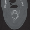
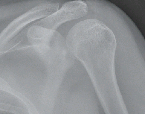 Figure 24A |
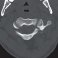
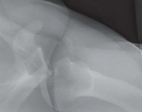 Figure 24B |
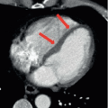
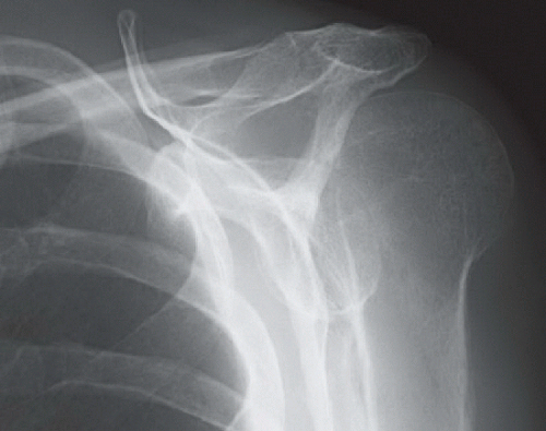 Figure 24C |
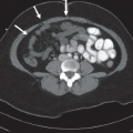
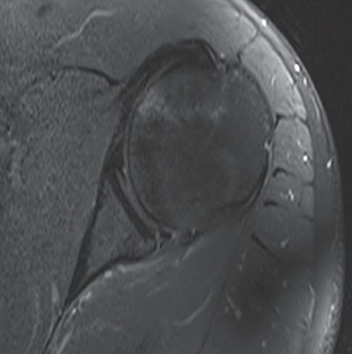 Figure 24D |
FINDINGS
An anteroposterior (AP) view of the left shoulder (Fig. 24A) with the proximal humerus in internal rotation demonstrates less than the usual amount of overlap between the humeral head and the glenoid expected for this view, sometimes described as a loss of the “half-moon overlap sign.” The axillary view (Fig. 24B) demonstrates posterior dislocation of the humeral head and a large triangular defect of the anterior aspect of the articular surface that is “engaged” with the posterior glenoid rim. A scapular-Y view in another patient (Fig. 24C) demonstrates posterior displacement of the humeral head in relation to the glenoid, also indicating a posterior dislocation. An axial T2-weighted fat-suppressed MR image in a third patient (Fig. 24D) demonstrates a posterior labral tear and a small contusion of the anterior humeral head articular surface, a reverse soft tissue Bankart and reverse Hill-Sachs lesion.
DIFFERENTIAL DIAGNOSIS
Posterior shoulder dislocation (On the AP view alone: Normal, posterior shoulder dislocation, pseudosubluxation).
DIAGNOSIS
Related posts:


Stay updated, free articles. Join our Telegram channel

Full access? Get Clinical Tree


