Primary Bone Neoplasms
Todd M. Blodgett, MD
Alex Ryan, MD
Joanna Costello, MD
Key Facts
Terminology
Chondrosarcoma
Ewing sarcoma
Osteogenic sarcoma
Imaging Findings
PET/CT improves accuracy of identification and localization of invasive disease
Crucial for determining therapeutic strategy
FDG PET superior for detection of osseous lesions; CT for lung lesions
Low FDG uptake and metabolic activity in cartilaginous tissue
FDG uptake increases with more aggressive histologic tumor types
High pre-treatment SUV sensitive for higher grade tumor
PET/CT superior to CT alone for detection of minimal malignant residue
Top Differential Diagnoses
Bone Metastases
Other Bone Tumors
Acute Leukemia
Benign Bone Lesions
Fracture
Abscess/Osteomyelitis
Bone Infarction
Paget Disease
Diagnostic Checklist
Best time to perform FDG PET or PET/CT is prior to therapy to establish inherent FDG activity
Many sarcomas, including primary osseous sarcomas, may not be FDG avid
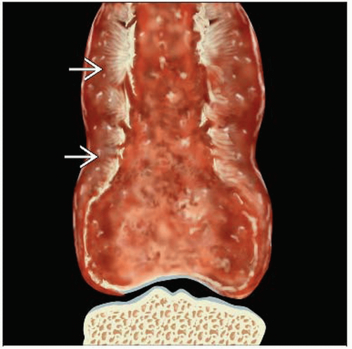 Graphic shows the typical appearance of a primary sarcoma of the femur  . . |
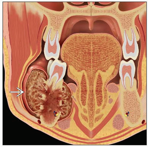 Coronal graphic shows the typical appearance of a mandibular osteosarcoma  . . |
TERMINOLOGY
Abbreviations and Synonyms
Chondrosarcoma
Ewing sarcoma
Osteogenic sarcoma, osteosarcoma, primary bone sarcoma
Definitions
Chondrosarcoma
Primary malignant tumor of bone
Produces hyaline cartilage leading to abnormal cartilage &/or bone
Ewing sarcoma
Primary malignant tumor of bone
Osteosarcoma
Primary malignant tumor of bone
Contains osteoid with osteoblastic differentiation
IMAGING FINDINGS
General Features
Best diagnostic clue
Chondrosarcoma
Mass with variable chondroid matrix
May have cortical disruption &/or soft tissue extension
FDG uptake tends to correlate with tumor grade
Ewing sarcoma
Permeative appearance ± extraosseous large soft tissue component adjacent to bone
Osteosarcoma
Heterogeneous metaphyseal mass
Increased FDG uptake with high grade osteosarcoma
May have characteristic starburst appearance
Location
Chondrosarcoma
Most common areas: Pelvis, femur, and humerus
Mostly in proximal aspect of long bones
Peripheral, periosteal, or central intraosseous locations
Ewing sarcoma
Most common in metaphysis or diaphysis of long bones
May arise in any bone
Osteosarcoma
Metaphyseal long bone
Distal femur
Size: Variable
Morphology
Ewing sarcoma
Obscured margins
Aggressive periosteal invasion
Osteosarcoma
Bone mass with destruction of bone elements
Cortical expansion
Large zone of transition
Chondrosarcoma
Endosteal scalloping
May have cortical destruction
Typically associated with a large soft tissue mass
Imaging Recommendations
Best imaging tool
FDG PET and PET/CT may be useful for staging, restaging, and response to therapy
Both have current insurance coverage limitations
CT Findings
Chondrosarcoma
Lytic mass with medullary cavity expansion
Variable amounts of calcification and chondroid matrix
Often have cortical thickening
± Soft tissue mass
± Endosteal scalloping
Ewing sarcoma
Commonly involves long bones (metaphysis or diaphysis)
Intramedullary mass ± involvement of the cortex
Permeative/“moth-eaten” appearance
Heterogeneous contrast enhancement
Periosteal reaction often described as “sunburst”
Large zone of transition
Soft tissue mass frequently present
Frequently metastasizes to lung, bone, and marrow
Osteosarcoma
Intramedullary mass
“Moth-eaten” appearance of osseous destruction
Indistinct borders
Wide zone of transition
Cortical break through
Most common in distal femur
Sunburst pattern of periosteal reaction
May have contrast enhancement
Nuclear Medicine Findings
Initial diagnosis
High grade tumors typically have intense FDG activity
FDG PET is sensitive for osteosarcoma and Ewing sarcoma
Chondrosarcoma shown to have low FDG uptake (average SUV of 4.5)
Histologic grade correlates well with SUV between high and low grade bone sarcomas
Staging
PET/CT improves accuracy of identification and localization of invasive disease
Crucial for determining approach to therapy
Invasion of adjacent structures can be determined
FDG PET superior for detection of osseous lesions
Useful for estimating percentage of tumor necrosis
PET/CT may offer additional biopsy localization information
CT more sensitive for lung lesions
Ewing sarcoma
Tend to be FDG avid
Small study reported overall sensitivity 96% and specificity 78%
PET/CT more sensitive than bone scan for osseous metastases
Degree of FDG uptake indicates disease stage and may have prognostic value
Chondrosarcoma
Low FDG uptake and metabolic activity in cartilaginous tissue
FDG uptake increases with more aggressive histologic tumor types
Cannot differentiate grade I chondrosarcoma from benign cartilage tumors
High pre-treatment SUV sensitive for higher grade tumor
High sensitivity for high grade mets
SUV may be as accurate as tumor grade for prognosis of overall survival
Restaging
PET/CT superior to CT alone for
Detection of minimal malignant residue
Distinction between vital and necrotic tumor regions
Improved detection and localization of recurrence in patients with implanted orthopedic devices
Response to therapy
Treated lesions may not change size after treatment, making metabolic imaging useful for evaluating response to therapy
Change in post-treatment maximum SUV correlates well with tumor response
Average SUV measurement prone to inconsistency based on tumor area chosen
Useful in Ewing sarcoma to assess response to induction chemotherapy
Less than 90% tumor necrosis response following pre-surgical treatmentRelated posts:

Stay updated, free articles. Join our Telegram channel

Full access? Get Clinical Tree


