Principles and physics of ultrasound imaging: simple terminology definitions

Chapter 2 Principles and physics of ultrasound imaging


simple terminology definitions


Absorption: This is the major cause of attenuation. Absorption occurs when ultrasound energy is lost to tissues by its conversion to heat. Higher frequency waves undergo greater absorption.


Acoustic impedance: A property of all substances and is equal to the product of the tissue density and the speed of sound. Comparatively speaking, two substances with greater differences in acoustic impedance produce stronger ‘echoes’ or reflected waves than two similar substances. Structures of different acoustic impedance (for example, gallbladder and gallstone) are easier to distinguish from one another than two structures of similar acoustic impedance (for example, liver and kidney).


Acoustic power: The rate of flow of energy through the cross-sectional area of the beam.


Acoustic waves: These are the vibrations that occur as a result of the rapid forward and reverse vibrations of the transducer, and which result in a number of longitudinal waves being transmitted. The transducer causes molecules in the medium through which it is passing to vibrate in a series of rhythmic, mechanical compressions (high-pressure regions) and rarefactions (low-pressure regions). These vibrations are commonly known as acoustic waves.


Acoustic window: An area of the patient that enhances ultrasound transmission and provides optimal scanning access to the area of interest. To improve image quality, ultrasound transmission should be as uniform as possible and areas which are likely to cause artifacts (such as ribs or bowel gas) should be avoided.


ALARA: An acronym for ‘as low as reasonably achievable’, referring to the principle of keeping power and exposure time to a minimum while acquiring the necessary clinical information.


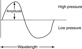
Amplitude: The height of a wave. The amplitude and intensity of sound represent the energy associated with the sound wave. The greater the amplitude or intensity, the more the energy, and the ‘louder’ the sound. Increasing the acoustic power will increase both the intensity and the amplitude (see Fig. 2.1).


Anechoic: Areas on the image showing no internal echoes, appearing dark or black on the image.


Artifacts: In the context of ultrasound, artifacts are echo signals whose displayed position on the image does not correspond to the actual position of a reflector in the body, or whose displayed brightness is not indicative of the reflecting or scattering properties of the region from which the echo originated. Artifacts are a result of the following programmed machine assumptions:


Attenuation: The process that occurs as a sound wave travels through a medium; it loses energy, and as a result, its intensity and amplitude decrease, and it becomes attenuated. Attenuation is proportional to the frequency of the sound wave and the distance that the wave travels. The higher the frequency and the further the wave travels, the greater the attenuation. Attenuation results from three main effects: absorption, reflection, and scattering.


Axial resolution: This refers to reflectors that lie along the axis of the ultrasound beam. This resolution is dependent upon the pulse length, which is equal to the product of the number of cycles in a pulse and the wavelength. If two reflectors along the axis of the ultrasound beam are separated by a distance longer than half the pulse length, they will appear as two separate reflectors. If the distance between the reflectors is less than the pulse length, they will appear as one reflector. Since the wavelength and frequency are inversely related, axial resolution is improved by increasing the frequency of the transducer. Thus, high-frequency transducers have better axial resolution.


Beam former: Provides pulse delay sequences to individual elements (an element consists of a piezoelectric crystal and its electrical connection) to achieve focusing of the ultrasound beam.


Cavitation: The pressure oscillations produced by sound can create gas bubbles from the air dissolved in tissue fluids. If the oscillations are rapid and intense, they can cause the bubbles to expand, contract, or collapse. The potential for cavitation is related to the acoustic pressure amplitudes produced by the ultrasound system. These amplitudes are reported by the manufacturer in the operator’s manual. Some scanning machines provide continuous assessment of the potential risk of bioeffects due to cavitation, by calculating the mechanical index (MI) for a given transducer in a particular mode during a scan. The MI is inversely proportional to the square root of the frequency; thus, as frequency increases, MI decreases.


Contact coupling: This can be either gel or liquid. Adequate coupling agent is needed to ensure that there is no air between the transducer and the skin.


Coronal plane: Divides the body into anterior and posterior sections, perpendicular to the sagittal.


Depth: The depth range or depth control varies the depth of the patient which is displayed on the image. The optimal depth is dependent upon beam penetration, which is determined by the transducer frequency.


Diffuse reflectors: These are also known as scatterers, and reflect sound in all directions. The brightness is not dependent on the angle of the incident beam.


Doppler shift:

Stay updated, free articles. Join our Telegram channel

Mar 10, 2016 | Posted by in ULTRASONOGRAPHY | Comments Off on Principles and physics of ultrasound imaging: simple terminology definitions

Full access? Get Clinical Tree

Get Clinical Tree app for offline access