Fig. 17.1
Bilateral nerve-sparing ablation
With cryoablation as the destructive energy force, this template can be employed either with or without the use of a cryoprobe positioned near one or both erectile nerves, which is warmed with pressurized helium. This template can also be achieved with high-intensity focused ultrasound (HIFU) wherein the energy is not directed laterally, near one or both neurovascular bundles.
It is well recognized that the posterior/lateral location is one of the common sites for prostate cancer. Canine data have raised concerns about cancer control when this template is employed [6]. Additionally, the location of the erectile nerves is now better described and known to exist in a broad swath of peri-prostatic tissue, not in a single, posterior lateral location [7]. Within the COLD registry, “nerve warming” cryoablation has not resulted in significant improvements in erectile function preservation.
Hemi-ablation
Hemi-ablation is the unilateral (hemisphere) destruction of all prostate tissue that is present to the left or right of the urethra (dictated by the laterality of the targeted cancer) and from apex to base, and anterior to posterior (Fig. 17.2).
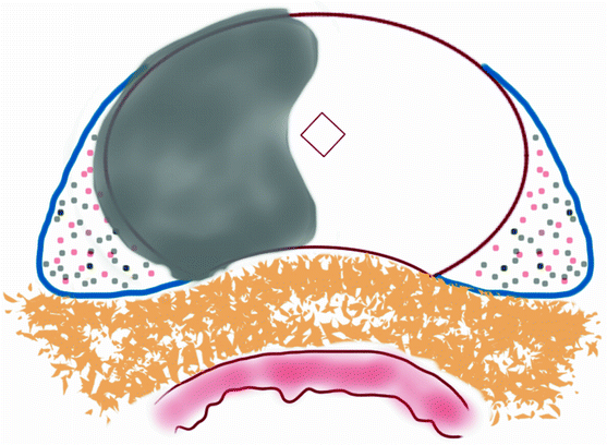

Fig. 17.2
Hemi-ablation
The theoretical benefit is to maximally preserve the neurovascular bundle contralateral to the treated, cancerous side of the prostate. This can be thought of as a unilateral nerve bundle preservation; however, the functional outcomes in the reports by Bahn and Onik where this template was employed are significantly better than is observed with unilateral nerve bundle preservation at radical prostatectomy [2, 3, 8].
Hockey-Stick Ablation (Anterior Three-Fourth)
Hockey-stick ablation is the extension of the hemi-ablation template across the midline only in the anterior region of the prostate, contralateral to the dominant cancer (Fig. 17.3).
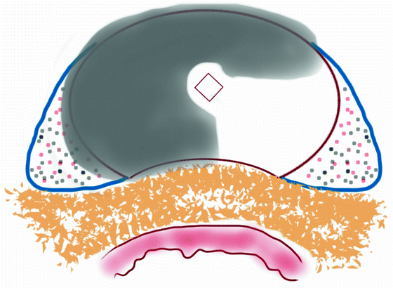

Fig. 17.3
Anterior hockey-stick ablation (right dominant with left wing)
Described first by Ward et al., this template has the theoretical benefits of treating potentially under-sampled, unrecognized cancers within the anterior region of the prostate contralateral to a dominant cancer that exists within the dominantly treated prostate hemisphere [9]. At a microscopic level, this template still provides preservation of the contralateral neurovascular bundle because of its delta shape in the periprostatic region; yet it provides a significant amount of prostate gland ablation, especially in a region of the prostate that is under-sampled by standard transrectal prostate biopsy. Currently, a large, prospective trial of focal cryoablation employs this template.
Posterior Three-Fourth Ablation
Posterior three-fourth ablation is the extension of the hemi-ablation to include the contralateral posterior region (Fig. 17.4). The template has the theoretical advantages of treating almost the entire prostate peripheral zone, which is the most likely location for cancer occurrence, while avoiding overlapping energy delivery to the urethra, which may cause urethral injury. However, the posterior peripheral zone of the prostate is better sampled than the anterior zone and thus contralateral significant tumors are more likely to be identified by standard prostate biopsy. Additionally, the delta-shaped neurovascular bundle is much more likely to receive damaging energy with this template, thus potentially negating the intent to preserve erectile function. However, in a prospective study that employed this template using HIFU as the ablative energy, it did result in decreased time of catheter bladder drainage than whole gland HIFU. Thus, this template may offer an improvement in the urinary morbidity profile, especially when erectile function is not adequate at baseline [10].
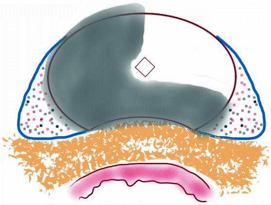

Fig. 17.4
Posterior hockey-stick ablation (right dominant with left wing)
Targeted Focal Therapy
Targeted focal therapy intends to minimize the collateral damage of surrounding normal prostate tissue and accurately target a volume of the prostate limited to the volume of the cancer (Fig. 17.5).
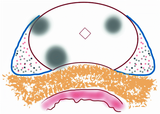

Fig. 17.5
Targeted focal therapy
Because our current ability to visualize the prostate cancer within the prostate gland is not at the level of sensitivity or specificity that exists in other organ sites, this template now requires a very accurate prostate mapping and the belief that this prostate mapping provides accurate enough information to create a three-dimensional model of the prostate and its contained tumors [11]. It is proposed that such definition of tumor location(s) may be achieved through a transperineal template-guided saturation biopsy schema followed by co-registering a three-dimensional image of the patient’s prostate with the histology [12


Stay updated, free articles. Join our Telegram channel

Full access? Get Clinical Tree


