Fig. 7.1
Discoloration of the digits and an ulcer on the ring finger (bandaged) went unnoticed by both the dialysis nurses and nephrologists in this patient referred for venous hypertension and inflammation at the cannulation sites
7.2.3 The Normal Vascular Access
A normal AVF is rare finding in the interventional suite. There should be a palpable thrill at the anastomosis, and arm elevation should empty the AVF. A thrill is caused by turbulent flow at the anastomosis as jets of blood flowing from a high pressure (130 mmHg) but smaller caliber artery are dumped into a larger more compliant but low pressure vein system.
7.2.4 The Flat Fistula
A flat arterialized vein accompanied by ecchymoses is very suggestive of an inflow problem likely at the anastomotic or juxta-anastomotic vein segment or afferent feeding artery (Fig. 7.2). The hematomas and ecchymoses are caused by multiple unsuccessful cannulations. Such AVFs are usually less than 1 year old.
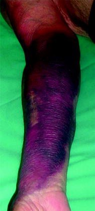
Fig. 7.2
Ecchymoses on the forearm arise from extravasation due to repeated failed cannulation of this flat radial–cephalic fistula harboring an anastomotic stenosis which should have been diagnosed and addressed earlier
7.2.5 The Hyperpulsatile Fistula
A tense AVF whereby the thrill is replaced by a water-hammer pulsatility indicates a venous outflow stenosis, often palpated as an indurated cord with a localized thrill (Figs. 7.3 and 7.4). The arterialized vein does not empty on arm elevation. There are usually inflammatory spots at the more recent cannulation points and collaterals can be seen.
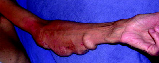
Fig. 7.3
This ulnar–basilic fistula, hyperpulsatile throughout its forearm course, has an outflow stenosis at the elbow resulting in aneurysmal degeneration
Fig. 7.4
(a) Typical presentation of inter-cannulation point stenosis in an old left brachial–cephalic fistula, manifesting as pulsatile aneurysmal arterial needling site and a spontaneously collapsed venous needling site aneurysm. (b) The clinical difference between the two aneurysms is exacerbated on arm elevation
7.2.6 The Falsely Normal Fistula
A combined inflow and outflow stenosis may give a false sense of a normal AVF on clinical examination. The palpation of subtle changes in the tone of the thrill along the arterialized vein and the auscultation of high-pitched thrill usually help clinch the diagnosis clinically.
7.2.7 The Inappropriately Needled Fistula
It is not unusual to find cannulation marks at the upper arm of a forearm AVF. This indicates either a stenosis or that the vein is too deep in the forearm (Fig. 7.5).
Get Clinical Tree app for offline access
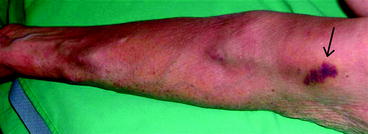
Fig. 7.5
This radial–cephalic fistula shows a well-developed vein a few centimeters from the anastomosis, which then disappears and reappears at the elbow. The ecchymosis on the upper arm (arrow
 Role of the Nephrologist, Interventional Radiologist, and Vascular Access in the Treatment of End-Stage Renal Disease
 Natural History of Vascular Access
Access Creation Strategy
Vascular Access Intervention Outcomes
Complications During and After Vascular Access Endovascular Procedures
Natural History of Vascular Access
Access Creation Strategy
Vascular Access Intervention Outcomes
Complications During and After Vascular Access Endovascular Procedures
 Angiography (Fistulography)


Related posts:
Role of the Nephrologist, Interventional Radiologist, and Vascular Access in the Treatment of End-Stage Renal Disease
Natural History of Vascular Access
Access Creation Strategy
Vascular Access Intervention Outcomes
Complications During and After Vascular Access Endovascular Procedures
Angiography (Fistulography)
Stay updated, free articles. Join our Telegram channel
Full access? Get Clinical Tree
